SMS 2011 December;17(2):53-57.
Published online 2011 December 30 |
| Copyright ⓒ 2010 Soonchunhyang Medical Science
|
| Frequency of Nasal Septal Perforation at the Suture Fixation Site of a Silastic Sheet Inserted during Nasal Surgery |
| Jang Yul Byun, Young Joo Park, Jae Yong Lee |
| Department of Otorhinolaryngology-Head and Neck Surgery, Soonchunhyang University Bucheon Hospital, Soonchunhyang University College of Medicine, Bucheon, Korea |
| Corresponding Author: Jae Yong Lee , Tel: +82-32-621-5448 , Fax: +82-32-621-5016 , Email: jyent@schmc.ac.kr
|

|
ABSTRACT
|

|
|
|
Objective: This study was performed to evaluate the frequency of nasal septal perforation at the suture fixation site of a silastic sheet inserted during nasal surgery. Methods: Seven hundred and twenty-one patients with silastic sheet insertion during common nasal surgeries were examined. The frequency of perforations and subjective symptoms of the patients were evaluated. Results: Nasal septal perforation at the suture fixation site occurred in seven patients (0.97%). In three patients, perforations occurred immediately after removal of the sheet, while four patients developed perforations 2 to 4 weeks later. In most cases, perforations were small and did not exceed 2 to 3 mm in diameter. No patient complained of nasal symptoms related to the septal perforation postoperatively. Conclusion: The frequency of the septal perforation at the suture fixation site of a silastic sheet was very low and subjective symptoms were absent. |
|
Keywords: Nasal septum; Perforation; Frequency; Silastic |
|
|
|
INTRODUCTION
|
|
|
Silastic is a silicone strengthened with a Dacron mesh that has been used in a variety of surgical fields for various purposes [1-10]. Silastic sheet may also be applied during common nasal surgeries, such as turbinate surgery, septal surgery, endoscopic sinus surgery (ESS), and polypectomy. Silastic sheet can be used to prevent synechia between turbinates and septum or lateral nasal wall, and to promote the healing in cases of septal mucosal injury. However, several complications related to silastic sheet insertion have been reported. These include obstructive symptoms as a result of displacement or migration, an increased rate of infection, and granulation tissue formation [1,11].
The most common causes of nasal septal perforation include external nasal trauma and septal surgery
[12]
. However, septal perforations may also occur at the suture fixation site of a silastic sheet due to necrosis of the septal cartilage and mucosa. We cannot conclude the definite cause of septal perforations because, to our best knowledge, no study has been conducted on the perforations associated with silastic sheet insertion. In addition, no study has reported the frequency of nasal septal perforation after silastic sheet insertion during nasal surgery. Thus, we evaluated the frequency of perforations at the suture fixation sites of silastic sheets and associated subjective symptoms of the patients. |
|
|
MATERIALS AND METHODS
|
|
|
This retrospective study examined the records of 721 patients who had silastic sheet insertion among 2,150 patients who had undergone nasal surgeries, such as turbinate surgery, septal surgery, ESS, and polypectomy from March 2004 to February 2010. Medical records for the type of surgery, surgical procedures, intraoperative findings, occurrence of septal perforations, and associated subjective symptoms of the patients were reviewed. The following cases were excluded from the study: 1) patients with bilateral injuries of septal mucosa at the corresponding area during septal surgery; 2) patients who had undergone excessive septal cartilage resection due to severe deviation of cartilage itself during septal surgery; 3) wide trimming of septal mucosa affected by diffuse polyposis; 4) patients with unilateral insertion of a silastic sheet; 5) patients with benign and malignant tumors of the nasal septum and nasal cavity; 6) pre-existing nasal septal perforation; 7) patients with perforations that occurred at non-suture sites; 8) patients with granulomatous disease or infectious conditions, such as syphilis and tuberculosis; and 9) patients who failed to follow up before the minimum 3-month period. This study was approved by the Institutional Review Board of Soonchunhyang University College of Medicine.
All surgical procedures were performed by the same surgeon. The followings were indications for silastic sheet application: 1) possibility of adhesion between the nasal septum and inferior turbinate after septal and/or turbinate surgery; 2) cases of septal mucosal injury during septal surgery; 3) to prevent obliteration of the olfactory cleft or synechia between the middle turbinate and nasal septum due to extended involvement of the middle and superior turbinate with polyposis; and 4) to prevent lateral synechia formation when an unstable, floppy middle turbinate occurred during ESS. When indicated, a soft, pliable, 0.03-inch-thick polymeric silastic sheet (Dow Corning, Midland, MI, USA) was shaped and placed in the nasal cavity. The silastic sheet was placed between the nasal septum and middle turbinate, and the size of the sheet was designed to cover most of the nasal septum. In case of indication 4, the silastic sheet was placed between the middle turbinate and lateral nasal wall. The fan-shaped silastic sheet was of sufficient size to cover the middle turbinate, approximately 1.5 to 2.0 cm from the caudal margin, to stabilize the middle turbinate
[10]
. The silastic sheet was placed in the nasal cavity under direct vision using a straight, 4-mm, 0° endoscope (Karl Storz, Tuttlingen, Germany) and then secured to the caudal portion of the nasal cartilaginous septum, using through-and-through mattress suture with 4-0 chromic catgut (Ethicon Inc., Somerville, NJ, USA). This procedure provided adherence of the silastic sheet to the septum, while leaving sufficient space to allow the patient to breathe comfortably through the nose.
Most patients visited our office once a week for 4 to 6 weeks, and once or twice a month for 2 months after the procedure. The silastic sheet was removed 10 to 14 days after nasal surgery, depending on the mucosal condition and healing process. Occurrence of nasal septal perforation and existence of associated subjective symptoms (nasal obstruction, crusting, bleeding, whistling, discharge, foul odor, and nasal pain) were evaluated and verified from medical records. |
|
|
RESULTS
|
|
|
| Among 721 patients who had a silastic sheet insertion, septal perforations at the suture fixation site occurred in seven patients (0.97%). The type of surgery, respective number of patients, and frequency of perforations are summarized in
Table 1
. For the convenience of description, we defined the anterior needle passage site through the nasal septum during through-and-through mattress suture as “anterior suture site” and posterior needle passage site as “posterior suture site.” Four patients had perforations at the anterior suture site
(Fig.1)
and two patients at the posterior site
(Fig.2)
. One patient had perforations at both the anterior and posterior sites. In three patients, perforations occurred immediately after removal of the sheet, while four patients developed perforations 2 to 4 weeks later. In most cases, perforations were small and did not exceed 2 to 3 mm in diameter. During follow-up, perforation size did not change in five patients. However, in two patients with an anterior perforation, the size increased to about 8 to 9 mm in diameter. In all of the patients with septal perforations, nasal mucosa around the perforation site healed well and no patient complain-ed of nasal symptoms related to the perforation. |
|
|
DISCUSSION
|
|
|
Silastic sheets have been used in various nasal surgeries, mainly for the prevention of synechiae and the promotion of mucosal heal-ing. Synechiae occurring after nasal surgeries often disturb postoperative management and are associated with recurrence of the nasal obstruction, eventually resulting in patient dissatisfaction. In patients who underwent ESS, synechia formation between the middle turbinate and lateral nasal wall can obstruct the outflow of the ethmoid, maxillary, and frontal sinus, leading to recurrent symptoms and necessitating synechia division or further surgery [1-10]. Additionally, in cases of septal and turbinate surgeries, synechiae between the nasal septum and inferior turbinate may occur and lead to recurrence of nasal symptoms. Silastic sheet insertion can prevent this complication by acting as a mechanical barrier. If septal mucosal injury occurs during septal surgery, a silastic sheet can accelerate the healing process by moistening and humidifying the injured site and by avoiding further trauma during postoperative care.
Several complications related with silastic sheet insertion have been reported, such as obstructive symptoms as a result of displace-ment or migration, the possibility of infection, and granulation tissue formation [1,11]. The application of silastic sheets does not always have beneficial effects. Some patients may complain of nasal pain due to irritation of nasal mucosa. Furthermore, some patients may complain of nasal obstruction due to crust formation and nasal discharge. However, this can be relieved by proper location of the sheet, frequent saline irrigation, and meticulous postoperative care. Crusting at the suture site can also be prevented by applying an ointment or emollient. Because this study was conducted in a retrospective manner, we could not evaluate patient discomfort numerically or statistically. However, placement of a silastic sheet for 10 to 14 days may not be a great burden or discom-fort to the patient, and a large number of patients did not notice the sheet in the nasal cavity.
As mentioned in the introduction, we cannot conclude the exact cause of septal perforations associated with silastic sheet insertion. However, in our opinion, the most possible and explainable cause of septal perforation is thought to be excessive tension when fixing the sheet to the nasal septum with suture material. This may compromise the blood supply, resulting in necrosis of the septal cartilage and mucosa. In all of our cases, through-and-through mattress suture was used for the fixation of a silastic sheet to the nasal septum. The suture material passed the nasal septum twice, anteriorly and posteriorly, and both sites received the similar tension during the fixation. Thus, our theory can be applied to both anterior and posterior perforations, and proper tension should be used when affixing the sheet. Too loose a knot will not cause the sheet to adhere to the septum and can cause swinging of the sheet and nasal obstruction during breathing. In contrast, excessively tight suturing causes pressure necrosis, as mentioned above. Four patients demonstrated delayed perforations. These patients developed crust on both sides of the suture site. In our opinion, this may slowly compromise the blood circulation and result in delayed perforations. We were always careful when affixing the sheet to the nasal septum with suture material. However, even when appropriate tension is applied, perforations may occur for reasons that are not apparent from the results of this study.
Our study showed that the frequency of nasal septal perforation at the suture site was very low (0.97%) and none of the patients with septal perforation complained of discomfort or symptoms postoperatively. This may be due to the small size of the perforation and complete mucosal healing of the perforation site.
In conclusion, the frequency of septal perforations occurring at suture fixation sites after applying silastic sheets was very low and there were no subjective symptoms. However, we suggest that reasonable criteria are necessary prior to silastic sheet insertion and appropriate tension should be applied while affixing a silastic sheet to the nasal septum. We believe that this report has value as the first study to evaluate the frequency of septal perforation occurring after application of silastic sheets in nasal surgery. |
|
|
|
FIGURES
|
|
|
|
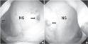
Fig.1
A patient who had undergone septal and turbinate surgery. Endoscopic views of the left (A) and right (B) nasal cavities show a small perforation (arrow) located at the anterior suture site 3 months postoperatively. IT, inferior turbinate; NS, nasal septum. |

|
|
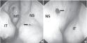
Fig.2
A patient who had undergone endoscopic sinus surgery and turbinate surgery. Endoscopic views of the left (A) and right (B) nasal cavities demonstrate a small perforation (arrow) located at the posterior suture site 3 months postoperatively. IT, inferior turbinate; MT, middle turbinate; NS, nasal septum. |
|
|
|
|
|
TABLES
|
|
|

|
Table.1
Type of surgery, number of patients, and frequency of perforations |
|
|
|
| |
|
|
REFERENCE
|
|
|
|
1.
|
Choi JS, Lee JH, Paik HJ. A silastic sheet found during endoscopic transnasal dacryocystorhinostomy for acute dacryocystitis. Korean J Ophthalmol 2006;20:65-9. |
|
|

|
|
2.
|
Park CH, Choi DJ, Lee JH, Hong SM, Kwon TK, Joung HH, et al. Endoscopic reduction of medial orbital wall fractures using the rolled silastic sheet technique. J Trauma 2009;66:1421-4. |
|
|
|
|
3.
|
Morrison AD, Sanderson RC, Moos KF. The use of silastic as an orbital implant for reconstruction of orbital wall defects: review of 311 cases treated over 20 years. J Oral Maxillofac Surg 1995;53:412-7. |
|
|
|
|
4.
|
Hwang K, Kita Y. Alloplastic template fixation of blow-out fracture. J Craniofac Surg 2002;13:510-2. |
|
|
|
|
5.
|
Banhiran W, Sargi Z, Collins W, Kaza S, Casiano R. Long-term effect of stenting after an endoscopic modified Lothrop procedure. Am J Rhinol 2006;20:595-9. |
|
|
|
|
6.
|
Schliephake H, Schmelzeisen R, Maschek H, Haese M. Long-term results of the use of silicone sheets after diskectomy in the temporomandibular joint: clinical, radiographic and histopathologic findings. Int J Oral Maxillofac Surg 1999;28:323-9. |
|
|
|
|
7.
|
Giancarlo H, Mattucci KF. Silastic sheet keel in laryngeal reconstruction. Laryngoscope 1985;95(9 Pt 1):1123. |
|
8.
|
Stefansson GM, Stilwell JH. Use of silastic sheet in Apert’s syndactyly. J Hand Surg Br 1994;19:248-9. |
|
|
|
|
9.
|
Michalevicz D, Chaimoff C. Use of a Silastic sheet for widening the abdominal cavity in the surgical treatment of diaphragmatic hernia. J Pediatr Surg 1989;24:265-6. |
|
|
|
|
10.
|
Lee JY, Lee SW. Preventing lateral synechia formation after endoscopic sinus surgery with a silastic sheet. Arch Otolaryngol Head Neck Surg 2007;133:776-9. |
|
|
|
|
11.
|
Groombridge C, McGuinness J. Interesting case: foreign body in the nose: an orbital silastic sheet had migrated into the nasal cavity. Br J Oral Maxillofac Surg 2005;43:56. |
|
|
|
|
12.
|
Kridel RW. Considerations in the etiology, treatment, and repair of septal perforations. Facial Plast Surg Clin North Am 2004;12:435-50, vi. |
|
|
|
|
|
|


