SMS 2012 June;18(1):9-11.
Published online 2012 July 17 |
| Copyright ⓒ 2010 Soonchunhyang Medical Science
|
| Is the Spinous Process of T7 Usually at the Same Level as the Inferior Tips of the Scapulae? |
| Mun Gyu Kim1, Si Young Ok1, Sang Ho Kim1, Se Jin Lee1, Sun Young Park1, Eun Hyo Koh1, Heon Yong Bae2, Kyung Yul Hur3
|
|
1Department of Anesthesiology and Pain Medicine, Soonchunhyang University Seoul Hospital, Soonchunhyang University College of Medicine; 2Department of Anesthesiology and Pain Medicine, Asan Medical Center, University of Ulsan College of Medicine;3Department of General Surgery, Soonchunhyang University Seoul Hospital, Soonchunhyang University College of Medicine, Seoul, Korea |
| Corresponding Author: Si Young Ok , Tel: +82-2-709-9302 , Fax: +82-2-790-0394 , Email: syok2377@naver.com
|

|
ABSTRACT
|

|
|
Objective: Appropriate placement of thoracic epidural catheter provides an adequate postoperative analgesia in chest and upper abdominal surgery. Usually, when thoracic epidural puncture is performed, both scapular lower tips and the thoracic (T)7 spinous process is assumed to be at the same horizontal level. The aim of this study is to identify the thoracic epidural puncture in the sitting position, with the neck flexed and arms crossed, may change the relationship between the thoracic vertebrae and the scapular lower tips.
Methods: One hundred patients with postoperative patient controlled epidural analgesia using thoracic epidural catheters were enrolled. It is presumed that the both scapular lower tips and T7 spinous process is at the equal level when performing thoracic epidural puncture. The actual insertion level of the Tuohy needle was examined by radiography when the patient was in the sitting position.
Results: Out of 100 patients, there were 62% that were in the same level as the scapular lower tips and T7 spinous process. However, 1% of the patients leveled at T4, 1% at T5, 25% at T6, 18% at T8, and 1% at T9.
Conclusion: When performing the thoracic epidural puncture under the sitting position, the relationship of the T7 and the scapular lower tips may change. The change of position of scapular lower tips varied among T6.82±0.70. Therefore, to be precise, it is advised to utilize C-arm guide when epidural puncture is carried out. |
|
Keywords: Scapular lower tips; Seventh cervical spinous process; Seventh thoracic spinous process; Surface anatomy landmark; Thoracic epidural puncture |
|
|
|
INTRODUCTION
|
|
|
Insertion of the epidural catheter congruent to the incisional dermatome provides optimal postoperative epidural analgesia and minimizing side effects. Use of high thoracic epidural analgesia for chest or upper abdominal surgery does not inhibit sympathetic nerve activity in the lower extremities, and relatively low incidence of urinary retention. Therefore when performing thoracic or upper abdominal surgery, first step for an effective postoperative epidural analgesia is to insert the epidural catheter at the adequate thoracic epidural level. Usually, when thoracic epidural puncture is performed, both scapular lower tips and the thoracic (T)7 spinous process are assumed to be at the same horizontal level
[1]
. But previous reports demonstrated that the use of horizontal line drawn between both scapular lower tips as surface anatomy landmark of T7 spinous process is inaccurate [2-4].
In the current study, we examine the change in the relation between scapular lower tips and thoracic vertebra when thoracic epidural puncture is performed at a sitting position, with the neck flexed and the arms crossed. |
|
|
MATERIALS AND METHODS
|
|
|
Following approval by the hospital research ethics committee, 100 adult patients over 20 years of age already scheduled for mastectomy, gastrectomy, hepatobiliary operation, Whipple procedure, and colectomy were enrolled in this study between August 2009 and May 2010. They were asked to join the study and written informed consent was obtained. Exclusion criteria included patients with body mass index >25, difficult thoracic spinous process palpation, obvious spinal deformity, spine operation history, or any contraindication to epidural anesthesia (history of allergic reactions to any local anesthetics, blood coagulation abnormality, and patient’s refuse to thoracic epidural puncture). Patient’s sex, age, height, and weight were recorded
(Table 1)
. And operation type were also recorded
(Table 2)
.
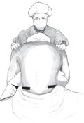
Horizontal line was drawn between to connect the scapular lower tips while the patient was in the sitting position, with the neck flexed and the arms crossed
(Fig.1)
. If the spinous process is palpated along the horizontal line drawn, it is predicted to be T7.
Epidural puncture was performed by setting T6-7 as a standard. In the case of mastectomy, T4-5 was used. For gastrectomy, hepatic resection, Whipple procedure, T6-7, or T7-8 was used. For colectomy T8-9 was used. Epidural puncture was performed with the midline approach. Then, the epidural space was identified using the loss-of-resistance technique with air. And, epidural catheter (Epidural catheter; Arrow International Inc., Reading, PA, USA) was inserted.
Anteroposterior and lateral X-ray view of thoracic spine had taken under Tuohy needle inserted. The diagnosis of the insertion level of epidural catheter and Tuohy needle was consulted with the radiologist. |
|
|
RESULTS
|
|
|
| There were 62% of the patients that resulted in accordance with the traditional assumption of having scapular lower tips and T7 in the same level. But 1% resulted in scapular lower tips in the position with T4, 1% with T5, 25% with T6, 10% with T8, and 1% with T9. The position of both scapula lower tips varied among T6.82± 0.70. |
|
|
DISCUSSION
|
|
|
The surface anatomy landmark of the most prominent cervical spinous process (cervical [C]7), horizontal line drawn between the scapular lower tip (T7), horizontal line between both iliac crests (lumbar [L]4, Tuffier’s line) was set to be the standard to perform the spinal or epidural puncture. However, previous studies demonstrated that during spinal anesthesia, even the commonly used Tuffier’s line did not accurately measure the L4 spinous process or the L4-5 intervertebral space [5-8]. The intercrestal line has been shown to coincide with the L4 spinous process or the L4-5 interspace in 78.6%, lower than L4 in 17.8%, and at the L3-4 interspace in 3.7% of patients
[5]
. And in other report when using Tuffier’s line landmark, anesthesiologists correctly identified a particular lumbar interspace only 29% of the time
[6]
. Kim et al.
[8]
recently published full-flexion of the lumbar spine is changed intercrestal line slightly from L4 in to L4-5.
There are two methods in determining the level of thoracic vertebra using the surface anatomy landmarks. One method is to count down the intervertebral space from C7, the most prominent cervical spinous process, to T7 in the caudal direction. Another method is to draw a horizontal line between the scapular lower tips and to determine the position of T7 by assuming that T7 spinous process lies between the horizontal line.
Bernards
[2]
described the first prominent spinous process is C7, most prominent spinous process is T1. And Lee et al.
[3]
reported in Korean adult, the first prominent spinous process was most commonly the C6 spinous process in both male and female subjects and most prominent palpable spinous processes were C7 in males and C6 in females in the largest number of subjects. Teoh et al.
[9]
reported the T7 spinous process was identified correctly 29% of the time with the C7 landmark and 10% of the time with the scapular landmark. Accuracy improved for T7±1 level to 78% and 42%, respectively. The mobility of the scapula may have contributed to its inaccuracy. And Haneline et al.
[10]
report the mean spinal level corresponding to the inferior angle of the scapula was midway between the T8-9 interspace and the upper T9 boby (range, lower T7 to upper T10).
In this study, patients had their hands in a contralateral knee position to flatten the back. The results of the presented investigation demonstrated the change of relationship between scapula lower tips and thoracic spine. The scapula lower tips differed in T6.82±0.70. Also, to perform a precise puncture, it is advised for anesthesiologist to use C-arm as a guideline. |
|
|
|
FIGURES
|
|
|
|
Fig.1
Sitting position with the neck flexed and the arms crossed for thoracic epidural puncture. |

|
|
|
|
TABLES
|
|
|

|
Table.1
Demographic data |
|
|

|
Table.2
Type of surgery |
|
|
|
| |
|
|
REFERENCE
|
|
|
|
1.
|
Kleinman W, Mikhail MS. Spinal, epidural, & caudal blocks. In: Morgan GE, Mikhail MS, Murray MJ, editors. Clinical anesthesiology. 4th ed. New York: McGraw Hill Medical Publishing Division; 2006. p. 300. |
|
2.
|
Bernards CM. Epidural and spinal anesthesia. In: Barash PG, Cullen BF, Stoelting RK, editors. Clinical anesthesia. 5th ed. Philadelphia: Lippincott Williams & Wilkins; 2006. p. 69 |
|
3.
|
Lee YB, Kim SY, Park JT, Han YK, Yoon KB. On the accuracy of cervicothoracic vertebral level determination by palpation of spinous processes. Korean J Anesthesiol 1999;37:608-12. |
|
4.
|
Stonelake PS, Burwell RG, Webb JK. Variation in vertebral levels of the vertebra prominens and sacral dimples in subjects with scoliosis. J Anat 1988;159:165-72. |
|
5.
|
Render CA. The reproducibility of the iliac crest as a marker of lumbar spine level. Anaesthesia 1996;51:1070-1. |
|
6.
|
Broadbent CR, Maxwell WB, Ferrie R, Wilson DJ, Gawne-Cain M, Russell R. Ability of anaesthetists to identify a marked lumbar interspace. Anaesthesia 2000;55:1122- |
|
7.
|
Furness G, Reilly MP, Kuchi S. An evaluation of ultrasound imaging for identification of lumbar intervertebral level. Anaesthesia 2002;57:277-80. |
|
8.
|
Kim JT, Jung CW, Lee JR, Min SW, Bahk JH. Influence of lumbar flexion on the position of the intercrestal line. Reg Anesth Pain Med 2003;28:509-11. |
|
9.
|
Teoh DA, Santosham KL, Lydell CC, Smith DF, Beriault MT. Surface anatomy as a guide to vertebral level for thoracic epidural placement. Anesth Analg 2009;108:1705-7. |
|
10.
|
Haneline MT, Cooperstein R, Young MD, Ross J. Determining spinal level using the inferior angle of the scapula as a reference landmark: a retrospective analysis of 50 radiographs. J Can Chiropr Assoc 2008;52:24-9. |
|
|
|


