INTRODUCTION
The introduction of endoscopic sphincterotomy in 1974 [1] led to major developments in therapeutic pancreaticobiliary endoscopy. As a result, endoscopic retrograde cholangiopancreatography (ERCP) has become a widely useful procedure for management of pancreaticobiliary disease.
Acute calculous cholecystitis is a complication of cholelithiasis. Cholelithiasis causes persistent obstruction of the gallbladder outlet because of impaction in the neck of the gallbladder, Harmann’s pouch, or cystic duct [2]. Exact diagnosis can be based on the history, physical examination, blood tests, and ultrasonography. There are several treatment modalities, including laparoscopic cholecystectomy, open cholecystectomy, medical therapy, and percutaneous cholecystostomy. However, endoscopic retrograde cholelithiasis removal using a stone basket is nearly impossible because of difficult anatomical approach.
In our patient, cholelithiasis was fortunately removed by ERCP. After the procedure, clinical finding of the patient showed dramatic improvement. We herein present a rare case of cholelithiasis successfully treated by retrograde endoscopic removal.
CASE REPORT
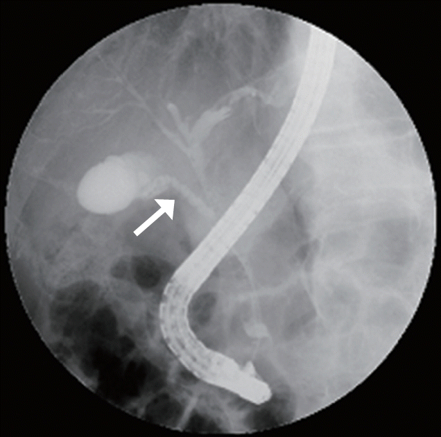
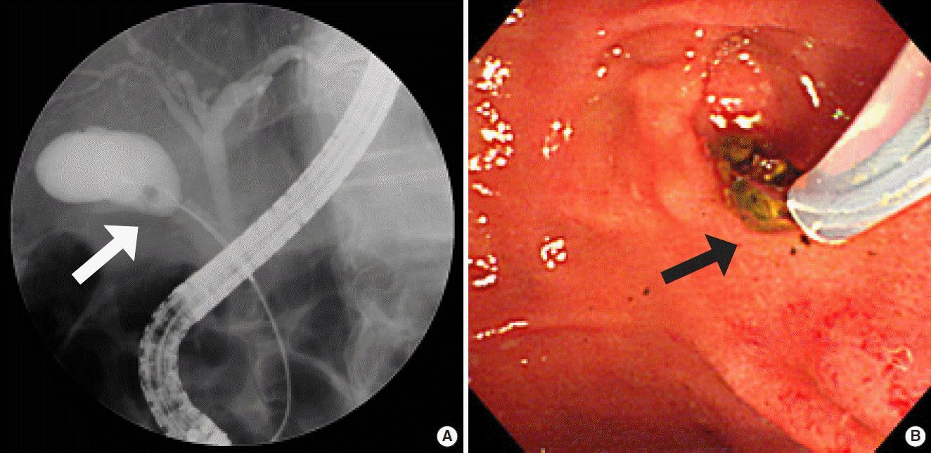
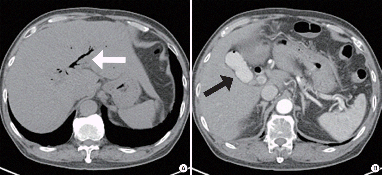
A 70-year-old male was admitted due to right upper quadrant pain and fever for 1 day duration. In a prior admission 1 year ago, he had already been treated with endoscopic retrograde choledocholithiasis removal. At admission, he was acutely ill-looking in appearance and body temperature was 38°C. Bowel sounds were normal and there was direct tenderness in the right upper quadrant, but no rebound tenderness. No abnormalities were detected during the remainder of the examination. Laboratory findings were as follows: white blood cell count 8,370/mm3, hemoglobin 16.3 g/dL, platelets 294,000/μL, aspartate aminotransferase 1,128 IU/L, alanine aminotransfere 512 IU/L, alkaline phosphatase 94 IU/L, total bilirubin 1.76 mg/dL, gamma-glutamyl transferase 174 IU/L, amylase 51 IU/L, lipase 131 IU/L, and C-reactive protein 0.32 mg/dL, with a normal prothrombin time/international normalized ratio. Ultrasonography showed a stone shadowing in the gallbladder with wall thickening. Computed tomography (CT) showed high-density material in a dependent position of the gallbladder and common bile duct (CBD). The clinical presentation suggested a diagnosis of acute calculous cholecystitis with choledocholithiasis and following ERCP. Endoscopic retrograde cholangiography showed a movable filling defect at the distal CBD and gallbladder, and cystic duct was short and extend in a straight line (Fig. 1). First, choledocholithiasis was removed using a stone basket, and then the basket was easily introduced into the gallbladder and the stone was removed successfully (Fig. 2). The patient’s condition then became noticeably better. Follow-up CT showed air-biliarygram with mild CBD dilatation, but there was no definite gallbladder and bile duct wall thickening or remnant stone (Fig. 3). He was discharged with oral antibiotics and has undergone follow-up in the outpatient department.
DISCUSSION
Acute calculous cholecystitis, which occurs as a complication of cholelithiasis, afflicts more than 20 million Americans annually [3]. Most patients with cholelithiasis are asymptomatic. Among those patients, 1% to 4% develops biliary colic annually [4], and approximately 20% of these symptomatic patients eventually develop acute cholecystitis if left untreated [5]. The Tokyo guidelines provide recommendations for management depending on the severity of acute cholecystitis [6]. Our patient’s presentation was consistent with mild acute cholecystitis, so that early laparoscopic cholecystectomy may be the treatment of choice. Considering the patient’s good condition after the procedure, it was not performed. However, laparoscopic cholecystectomy would be necessary in case of recurrence.
In the Western world, most stones in the CBD arise from the passage of cholelithiasis into the CBD. Stones in the common duct occur in 10% to 15% of patients with cholelithiasis. Coexistent cholelithiasis and choledocholithiasis occur more frequently in elderly, Asian patients and in patients with chronic inflammatory conditions such as primary sclerosing cholangitis, acquired immunodeficiency syndrome, parasite infection, and, probably, hypothyroidism.
In our case, coexistent cholelithiasis and choledocholithiasis were diagnosed by ERCP. A stone basket was easily introduced into the gallbladder and CBD, then stones were fortunately removed. However, endoscopic retrograde cholelithiasis removal is known to be difficult because of the anatomical approach. In one study cystic and common hepatic ducts in 524 persons were evaluated retrospectively in cholangiograms obtained for a variety of indications. The cysticohepatic junction was adequately visualized in 70% of cases. Medial junctions were noted in 18% and a spiral configuration in 32%, both more common than reported. An 11% occurrence of parallel duct systems was less frequent than expected. In 10% of patients, the cystic duct entered the hepatic duct in the distal third of the extrahepatic biliary tree. Our patient’s cystic duct had a relatively straight configuration, so that removal of cholelithiasis was easier. Thus, understanding of these anatomical variations is considered important for therapeutic endoscopic procedures.
Since development of laparoscopic cholecystectomy in 1987, it has become the treatment of choice for symptomatic cholelithiasis [7]. Although it has been widely used, occasional complications after surgery have been limited to elderly patients with comorbid conditions. In some literature, bile duct injuries, which occurred in 0.6% of patients during surgery, were particularly troublesome. The mean rate of bowel and vascular injuries, the most lethal complications, was 0.14% and 0.25% of cases [8]. For these reasons, there has been significant effort for cholelithiasis extraction for patients who are not suitable for surgery. Percutaneous cholecystostomy was first described by Radder in 1980, avoids the use of general anaesthesia, invasive surgery and attendant risk while effectively managing gallbladder disease in critically ill elderly patients. The reported technical success rate is 98% to 100% with few procedure related complications. The most commonly reported minor complications include percutaneous catheter dislodgement or blockage, and major complications include bleeding, biliary injury resulting in bile leakage, intestinal perforation or pneumothorax [9]. Endoscopic transpapillary cystic duct cannulation was first described in 1984. According to one report 50 consecutive patients with a variety of pancreaticobiliary conditions were studied prospectively. In 74%, the cannula could be inserted selectively into the cystic duct [10]. Direct cholelithiasis dissolution by instilling solvent has also been reported. Using retrograde endoscopic placement of a nasobiliary catheter into the cystic duct, solvent could be delivered to stones within the gallbladder. Endoscopic transpapillary pernasal gallbladder drainage and endoscopic gallbladder stenting have recently been reported to show a clinically favorable response for acute cholecystitis. In addition, ERCP utilizing the spyglass cholangioscopy system, enabling direct visualization of the cystic duct and gallbladder, can access the cystic duct through which the gallbladder was ultimately decompressed, via biliary stent placement and cholelithiasis irrigation.
Endoscopic retrograde cholelithiasis removal using a stone basket is nearly impossible due to difficult anatomical approach. It could be used for extraction of cholelithiasis and could also be an alternate procedure like cholelithiasis dissolution, gallbladder stent drainage, and cholelithiasis irrigation. However, more research is needed to determine usefulness and limitation of endoscopic retrograde cholelithiasis removal using a stone basket. In addition, studies of endoscopic instruments such as the spyglass cholangioscopy system and stone extraction accessories are also necessary.
In this case, basket is fortunately easy to pass through the cystic duct and gallstones were removed. But in most cases it can lead to serious complications, such as strangulation, perforation of the gallbladder, and can lead to a gallbladder surgery. In this case, the patient was fortunately removed the gallbladder stones by ERCP and had good prognosis. We report this unusual case.


