INTRODUCTION
Thoracic splenosis is a rare condition resulting from splenic and diaphragmatic injury. Patients are usually asymptomatic, and so it is often discovered incidentally while computed tomography (CT) scan or magnetic resonance imaging (MRI) is performed for other reasons. Because disease is rare, patients often undergo an invasive diagnostic procedures including thoracotomy. The combination of imaging findings and clinical history make it avoid unnecessary invasive diagnostic procedure to confirm. Here we report a case of thoracic splenosis diagnosed without invasive procedure; additionally, we review the relevant literature.
CASE REPORT
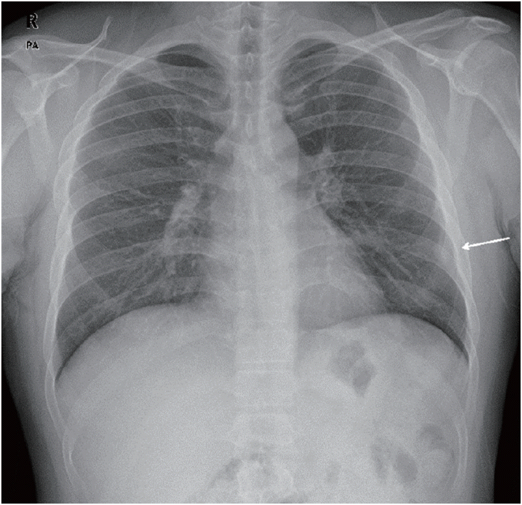
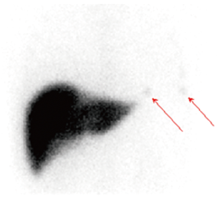
A 35-year-old man was referred to Soonchunhyang Cheonan Hospital for evaluation of incidental left diaphragmatic consolidation on chest X-ray (Fig. 1). He had been in good general conditions and had a 15-pack-year history of smoking. He did not have any symptoms, physical examination was unremarkable, and all laboratory tests were within normal ranges. From further medical history obtained, he had undergone closed thoracostomy and splenectomy following diaphragm rupture and splenic trauma from a traffic accident when he was fifteen years old. A chest radiograph demonstrated 1.5-cm sized subdiaphragmatic nodular opacity in left lower lung field. A chest CT demonstrated multiple tiny to 2-cm sized homogenously well-enhancing nodular lesions along pleura, extrapleural space, diaphragm, and anterior mediastinum of left side (Fig. 2). Given the information of trauma history and CT findings, we suspected thoracic splenosis and performed a 99mTc sulfur colloid liver scan (Fig. 3) which demonstrated multiple small foci of uptake in the left hemithorax, consistent with thoracic splenosis. This patient had been followed up at our outpatient clinic without further invasive evaluation or treatment for the thoracic splenosis.
DISCUSSION
In 1896, Albrecht first report a case of splenosis, and the thoracic splenosis was first reported by Bernard Shaw and Shafi [1] as an autopsy finding in 1937 [2]. Splenosis can develop in everywhere of the body and the most frequent organ is peritoneal cavity. Thoracic cavity is the most common organ for the extraperitoneal splenosis because of the anatomic boundaries of the abdominal and thoracic cavities [3]. The incidence of splenosis after splenic rupture is 76% and after diaphragmatic rupture is up to 18% [4]. Thoracic splenosis is usually due to simultaneous rupture of the spleen and the diaphragm as a result of trauma, which allows the implantation of splenic tissue in the left pleural cavity [2,5]. A few reported cases without diaphragmatic rupture have been attributed to the passing of splenic tissue across the diaphragm into the thoracic cavity [6].
While abdominal splenosis can make gastric symptoms such as abdominal pain or bowel obstruction, thoracic splenosis is commonly asymptomatic, although pleuritic pain or hemoptysis may present. Thoracic splenosis has been often recognized incidentally as solitary or multiple pleural or subpleural nodules on CT scan or MRI for other reasons several years after trauma. Therefore, without the clinical information, these nodules may be mistaken for pleural masses including pleural metastasis, lymphoma, or malignant mesothelioma.
Previously, many cases of thoracic splenosis had been diagnosed through invasive procedure as percutaneous biopsy, thoracotomy, or video-assisted thoracotomy when the splenosis was not considered at first. However, it has been now well-established that the diagnosis of thoracic splenosis can be confirmed by radionuclide imaging study without unnecessary biopsy or surgical procedures. Radionuclide imaging studies are usually obtained by scintigraphy using Tc-99m sulfur colloid, indium-III-labeled platelets and Tc-99m-labeled heat-damaged red blood cell (RBC) [7]. In Korea, only 1 thoracic splenosis case reported in the literature has been diagnosed in this way without resorting to invasive techniques to confirm a pathologic diagnosis [8]. The process is benign in most asymptomatic cases and treatment is generally conservative and wait-and-see approach that includes careful clinical and radiologic follow-up [9].
In conclusion, thoracic splenosis should be considered in the differential diagnosis of asymptomatic patients with multiple, left-sided pleural-based nodules and previous history of thoracoabdominal injury and splenectomy. Once suspected, the diagnosis of thoracic splenosis can be confirmed noninvasively using Tc-99m-labeled heat-damaged RBC scan, and unnecessary invasive procedure can be avoided.