SMS 2011 December;17(2):65-71.
Published online 2011 December 30 |
| Copyright ⓒ 2010 Soonchunhyang Medical Science
|
| Direct Comparison between Brachial Pressure Obtained by Oscillometric Method and Central Pressure Using Invasive Method |
| Sang-Ho Park, Seung-Jin Lee, Jae Yun Kim, Min Jeong Kim, Ji Yeon Lee, A-Ra Cho, Hyeok-Gyu Lee, Se-Whan Lee, Won-Yong Shin, Dong-Kyu Jin |
| Department of Internal Medicine, Soonchunhyang University Cheonan Hospital, Cheonan, Korea |
| Corresponding Author: Seung-Jin Lee , Tel: +82-41-570-3671 , Fax: +82-41-574-5762 , Email: drlsj@schca.ac.kr
|

|
ABSTRACT
|

|
|
|
Objective: The importance of central blood pressure evaluation for cardiovascular risk stratification has been emphasized. The aim of this study is to evaluate whether brachial blood pressure obtained by the oscillometric method accurately reflects central blood pressure. Methods: The subjects consisted of 84 consecutive patients with suspected coronary artery disease who underwent cardiac catheterization. Central blood pressure was invasively measured in the origin of the left subclavian artery by using the fluid-filled system, and at the same time, brachial blood pressure in the left upper arm was measured by the oscillometric method. Results: No significant difference was found between central systolic pressure and brachial systolic pressure (144.49±18.84 mmHg vs. 142.44±14.96 mmHg, P=0.063). Bland-Altman analysis accounted for only a small bias of +2.25 mmHg, and the limits of agreement were 24.15 mmHg and -19.65 mmHg. Central diastolic pressure was significantly lower than brachial diastolic pressure (75.80± 8.74 mmHg vs. 86.70±10.48 mmHg, P<0.001). Bland-Altman analysis showed a significant bias of -5.45 mmHg, and the limits of agreement were 2.83 mmHg and -13.73 mmHg. Conclusion: These results indicate that central systolic pressure can be directly estimated from brachial systolic pressure using the noninvasive oscillometric method and observed biases seem to remain within the practical range. However, use of the brachial diastolic pressure and pulse pressure measured by the noninvasive oscillometric method is doubtful in clinical practice because of their large biases. |
|
Keywords: Blood pressure; Oscillometry; Coronary artery disease; Bias |
|
|
|
INTRODUCTION
|
|
|
| Central blood pressure, indicating signs for the direct cardiac afterload, compared with peripheral blood pressure reflect more accurately the left ventricular myocardium, coronary and cerebrovascular events
[1]
. The importance of central blood pressure evaluation for cardiovascular risk stratification has been emphasized
[2]
. Various methods have been developed to measure central blood pressure. A previous study showed that radial systolic arterial pressure gives a poor estimate of the actual pressure in the ascending aorta, while the radial mean and diastolic arterial pressure are reliable
[3]
. Some investigators reported that central pressure waveform can be precisely generated from the radial pressure waveform obtained by applanation tonometry with the use of general transfer function
[4-6]
, whereas, others reported that a significant underestimation of central systolic pressure and pulse pressure was caused by radial pressure waveform [7-9]. In clinical settings, brachial arterial blood pressure is commonly used. Brachial systolic and diastolic pressures measured with a cuff-type sphygmomanometer may give better estimates of central systolic and diastolic pressures
[7-10]
. On the other hand, several investigators reported that there is a debate on the biases of accuracy in the cuff-type sphygmomanometer when used to measure systolic and diastolic brachial pressures [11,12]. The purpose of the present study is to evaluate whether brachial blood pressure obtained with a cuff-type sphygmomanometer actually reflects central blood pressure measured by the invasive method. |
|
|
MATERIAL AND METHODS
|
|
|
1. Study Patients
Among the patients who underwent coronary angiography due to chest pain from January 2010 to July 2010 at Soonchunhyang University Cheonan Hospital in South Korea, 84 patients who show-ed no significant stenosis (less than 50% of the reference diameter) and were negative for ergonovine provocation test, were enrolled in this study. The patients consisted of 27 males and 57 females. This study excluded the patients with: brachial and central systolic pressure ≥200, significant valvular heart disease, significant cardiac arrhythmia, an ejection fraction less than 50%, significant valvular heart disease, a serum creatinine above 1.5 mg/dL, a tortuous aorta, heart rate less than 50 beat per minute or more than 100 beat per minute, and diabetes mellitus. In all patients, pulsation of the left brachial artery was overt at their antecubital fossae. The study was approved by the local ethics committee, and written informed consent was obtained from all subjects.
2. Measurement of the Brachial Blood Pressure, Central Blood Pressure and Pulse Wave Velocity
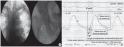
Shortly after diagnostic coronary angiography, a 5-F Judkins Right (JR) catheter was placed just distal to the origin of the left subclavian artery. Central blood pressure was measured at this site with a 5-F JR catheter using a fluid-filled system. While obtaining central blood pressure, brachial blood pressure in the left upper arm was simultaneously measured with a validated and calibrated automated cuff-type sphygmomanometer (H9000SE, Mennen Medical, Israel) using the oscillometric method. The measurement of upper arm pressure was made only once while obtaining central blood pressure. Pulse wave velocity (PWV) measurement and surface electrocardiography were simultaneously performed at a speed of 100 mm/sec via the JR catheter, and a 6-F sheath was placed in the right iliac artery using the fluid-filled system
(Fig. 1A)
. PWV was defined as distance/pulse wave transition time. The distance was defined as the length of the catheter exposed outside and the length of the sheath (12 cm) subtracted from the total length of the catheter (100 cm). The pulse wave transition time was obtained by subtracting the transition time of the descending aorta from that of the right iliac artery using the foot-to-foot method and measuring the interval between the beginning of QRS wave and the starting position of the first increase in pulse wave on surface electrocardiography
(Fig. 1B)
. To minimize errors in measuring transition time, we used the mean value measured from 3 consecutive pulse waves.
3. Statistical Analysis
Data are presented as means±SD. A value of P<0.05 was considered statistically significant. All statistical analyses were performed using SPSS ver. 14.0 (SPSS Inc., Chicago, IL, USA). Differences between central and brachial blood pressures were tested using the paired Student’s t-test. Bland-Altman analysis
[13]
was applied to the evaluation of agreement between the 2 methods of pressure measurement. The Pearson’s correlation was used to identify the correlation between central blood pressure, brachial blood pressure and PWV. |
|
|
RESULTS
|
|
|
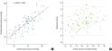
| The mean age of the patients was 57.87±13.86 years. No significant difference was found between central systolic and brachial systolic pressures (144.49±18.84 mmHg vs. 142.44±14.96 mmHg, P=0.063)
(Table 1)
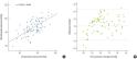
. There was a significant correlation between the 2 parameters (r=0.828, P<0.001) (Fig. 2A). Bland-Altman analysis accounted for only a small bias of +2.25 mmHg, and the limits of agreement were 24.15 and -19.65 mmHg (Fig. 2B). Central diastolic pressure was significantly lower than brachial diastolic pressure (75.80±8.74 mmHg vs. 86.70±10.48 mmHg, P< 0.001)
(Table 1)
. There was a significant correlation between the 2 parameters (r=0.643, P<0.001)
(Fig. 3A)
. Bland-Altman analysis showed a significant bias of -5.45 mmHg, and the limits of agreement were 2.83 mmHg and -13.73 mmHg
(Fig. 3B)
. Central pulse pressure was significantly greater than brachial pulse pressure (68.64±15.85 mmHg vs. 55.86±12.34 mmHg, P<0.001)
(Table 1)
. There was a significant correlation between the 2 parameters (r=0.726, P<0.001)
(Fig. 4A)
. Bland-Altman analysis revealed a significant, large bias of +12.79 mmHg, and the limits of agreement were 34.67 mmHg and -9.09 mmHg
(Fig. 4B)
. PWV indicating arterial stiffness was significantly correlated with central systolic pressure (r=0.464, P<0.001), central pulse pressure (r= 0.556, P<0.001), brachial systolic pressure (r=0.440, P<0.001) and brachial pulse pressure (r=0.512, P<0.001). PWV was also significantly correlated with age (r=0.512, P<0.001). The mean±SD of PWV was 10.05±3.03 m/sec. PWV was higher in the older and hypertensive groups
(Table 2)
. In the normotensive group (central systolic pressure<140 mmHg, n=32), a significant difference was found between central systolic and brachial systolic pressures (125.06±8.83 mmHg vs. 129.44±8.38 mmHg, P=0.006)
(Table 3)
. Bland-Altman analysis showed a significant, small bias of -4.38 mmHg, and the limits of agreement were 12.30 mmHg and -21.06 mmHg. However, in the hypertensive group (central systolic pressure≥140 mmHg, n=52), central systolic pressure was significantly higher than brachial systolic pressure (156.77±11.82 mmHg vs. 150.44±12.25 mmHg, P<0.001)
(Table 3)
. Bland-Altman analysis showed a significant, large bias of 6.33 mmHg, and the limits of agreement were 27.13 and -14.47 mmHg. In the younger group (age<60, n=46), no significant difference was found between central systolic and brachial systolic pressures (139.00±18.16 mmHg vs. 138.24±12.21 mmHg, P>0.05)
(Table 4)
. Bland-Altman analysis showed a small bias of 0.76 mmHg, and the limits of agreement were 23.54 and -22.02 mmHg. However, in the older group (age≥60, n=38), central systolic pressure was higher than brachial systolic pressure (151.58±17.49 mmHg vs. 147.53±16.49 mmHg, P=0.02)
(Table 4)
. Bland-Altman analysis showed a significant, small bias of 4.05 mmHg, and the limits of agreement were 24.54 and -16.43 mmHg. |
|
|
DISCUSSION
|
|
|
The gold standard for the measurement of arterial pressure is direct intra-arterial measurement with a catheter. However, this technique is neither practical nor appropriate for repeated measurements in nonhospitalized patients or asymptomatic individuals at a large-scale public health screening. Instead, the indirect measurement method is commonly used. By using the tonometric sensor to measure blood pressure was traditionally. However, this method was limited in technical accuracy, obesity and reproducibility of measurements. Thus, using relatively simple oscillometric method to measure peripheral blood pressure became a way to develop
[14]
. It has been known for more than 50 years that there are considerable differences in brachial pressure between cuff measurements and invasive measurements, especially in older subjects [15-17]. Smulyan et al.
[18]
reported that an inaccuracy of the oscillometric cuff mothod for measuring pressures from the upper arm appears to be a limiting factor. O’ Rourke and Adji
[12]
also stated that there is a debate on the biases of accuracy of the cuff-type sphygmomanometer to measure systolic and diastolic brachial pressures. The oscillometric method is based on detecting oscillations on the lateral wall of the occluded artery as the cuff is deflated. The oscillations begin at approximately the level of systolic pressure and reach their greatest amplitude at the level of mean arterial pressure. Diastolic pressure is a derived value. Perloff et al.
[11]
reported that systolic blood pressure measurement by this me-thod is accurate, but diastolic pressure, which is derived from systolic and mean pressures, may not be accurate. Some previous stu-dies showed that brachial diastolic pressure measured by the cuff-type oscillometric method was higher than directly recorded aortic central diastolic pressure
[19]
. The result of the present study corresponds well with those of earlier studies which reported that brachial diastolic pressure measured by the oscillometric method does not correctly reflect central diastolic pressure. On the other hand, Finnegan et al.
[20]
demonstrated in a study involving a volunteer group of 57 subjects (mean age, 68.6 years) that cuff-type measurements significantly underestimate systolic pressure by 5 mmHg compared to catheter measurements. However, Davies et al.
[7]
reported an important finding that peripheral systolic blood pressure measurements using a cuff-type automated sphygmomanometer from the left upper arm overestimated invasive measurements of systolic blood pressure in the ascending aorta only by an insignificant value of 3.36±10.47 mmHg. Cloud et al.
[9]
also reported that noninvasive brachial systolic pressure underestimates the catheter measured peak aortic pressure by only an insignificant value of 1.9 mmHg. Likewise, the present study showed that there was no significant difference between central systolic pressure measured in the aortic arch and brachial systolic pressure obtained from the left upper arm by the oscillometric method in patients with suspected coronary artery disease. Also, the observed biases seemed to remain within the practical range. The peak systolic pressure is greater in the upper arm than in the aorta due to amplification
[21]
. This amplification is related to the intensity of wave reflection, the difference in stiffness between central and distal arteries, the blood pressure level and PWV [22-25]. Our study subjects consisted of patients with suspected coronary artery disease aged 57.87±13.86 years, and they showed a high PWV (10.13 ±3.06 m/sec). We presumed that pressure waves were less amplified in our study, which resulted in no significant difference between central and brachial systolic pressures. With increasing age and degree of hypertension, arteriosclerosis or atherosclerosis, arterial wall thickness increases and presents functional and structural abnormalities [26,27]. These alterations predominate much more in the central elastic arteries than in the distal ones, and thus can generate an increase in central pressure pulse [21,27,28]. With increasing age and arterial stiffening, the modifications in the contour and amplitude of aortic pressure pulse are associated with a decrease in pulse amplification between central and peripheral arteries [26,29]. In our study, the mean value of central systolic pressure was higher than that of brachial systolic pressure (151.58 mmHg vs. 147.53 mmHg) in the older group (age≥60 years). Also, in the hypertensive group (central systolic pressure≥140 mmHg), the mean value of central systolic pressure was higher than that of bra-chial systolic pressure (156.77 mmHg vs. 150.44 mmHg). As mentioned above, the finding that central systolic pressure is higher in the older and hypertensive groups seems to be associated with an increase in PWV and augmentation of aortic pressure caused by the early return of reflected waves from the lower body and a decrease in the pulse amplification. On the other hand, since our study was done using a fluid filled catheter system, the results could be due to the overshooting of the systolic blood pressure. Assessment according to the USA Association for the Advancement of Medical Instrumentation SP10 criteria
[30]
for arterial pressure measurement with an automated sphygmomanometer requires a mean difference of ±5 mmHg and SD ±8 mmHg (2 SD, ±16 mmHg) from the reference standard. In our study, although pressure measurements were made at 2 different sites, the mean difference in systolic pressure between the aortic arch and left upper arm remained less than ±5 mmHg. However, the mean difference in diastolic pressure and the standard deviation of the differences between paired measurements in systolic pressure exceeded the above criterion. The relative large standard deviations of the differences between paired measurements of aortic and brachial systolic pressures are considered to be caused by the poor reproducibility of the single measurement from the upper arm with a cuff-type sphyg-momanometer [12,18]. In any case, single measurement of blood pressure with a sphygmomanometer and unusual circumstances in a catheter laboratory causes large errors
[31]
. Also, the oscillometric technique is particularly vulnerable to error in certain clinical circumstances, such as patients with arrhythmias and elderly patients with stiff arteries due to atherosclerosis
[32]
. The impact of human error on blood pressure measurement is a well-described and substantial problem. Human errors include inaccurate cuff selection and application, incorrect cuff positioning, inadequate rest period, rapid cuff deflation rate, poor observer concentration, digit bias and lack of repeated measurements. Use of automated devices rectifies some but not all of these problems
[33]
. However, if we had made measurements from the upper arm by oscillometric method as many times as practically possible to obtain the average values, systolic central pressure could have been replaced by systolic pressure obtained from the upper arm in a study group with less pressure amplification. Our study is a cross-sectional, observational one. Also, it has a limitation stemming from its small sample size.
In conclusion, the results of the present study suggest that brachial blood pressure obtained by the oscillometric method can pre-cisely reflect central blood pressure (central systolic pressure) measured by the invasive method. The biases of systolic pressure between 2 different sites seem to remain within the practical range. However, although central diastolic pressure and pulse pressure were well correlated with those measured by the noninvasive oscillometric method, there was some doubt in its use at clinical settings because of their large biases. Central systolic pressure seems to be more affected with age and degree of hypertension than brachial systolic pressure and higher in older and hypertensive subjects. Because the random variation of single pressure measurement in the upper arm may be large and unacceptable for clinical purposes, measurement by the oscillometric method should be repeated as many times as possible. |
|
|
|
FIGURES
|
|
|
|
Fig.1
(A) 5-F Judkins catheter was placed just distal to the origin of the left subclavian artery to calculate the distance between the pulse waves of the aorta and right iliac artery. (B) The length of the sheath (12 cm) exposed outside the sheath was subtracted from the total length of the catheter (100 cm). |

|
|
Fig.2
(A) There was a significant correlation between central systolic pressure measured at the ascending aorta with invasive method and the systolic pressure at left brachial artery obtained by oscillometric method. (B) Bland-Altman plots for systolic pressure measured by the the both sites. The analysis showed only a small bias of +2.25 mmHg, and the limits of agreement were 24.15 mmHg and -19.65 mmHg. Difference=central pressure-brachial pressure. Average=(central pressure+ brachial pressure)/2. |
|
|

|
Fig.3
(A) There was significant correlation between the central diastolic pressure measured at the ascending aorta with invasive method and the diastolic pressure at left brachial artery obtained by the oscillometric method. (B) Bland-Altman plots for diastolic pressure measured at both sites. This analysis showed a significant but relative small bias of -5.45 mmHg, and the limits of agreement were 2.83 and -13.73 mmHg. Difference=central pressure-brachial pressure. Average=(central pressure+brachial pressure)/2. |
|
|
|
Fig.4
(A) There was a significant correlation between central pulse pressure measured at the ascending aorta by the invasive method and pulse pressure at the left brachial artery obtained by the oscillometric method. (B) Bland-Altman plots for systolic pressure measured at both sites. This analysis showed a significant, large bias of 12.79 mmHg, and the limits of agreement were 34.67 and -9.09 mmHg. Difference=central pressure-brachial pressure. Average=(central pressure+brachial pressure)/2. |
|
|
|
|
|
TABLES
|
|
|

|
Table.1
Comparison between brachial and central pressures |
|
|

|
Table.2
Comparison of the mean values of PWV according to age and central systolic pressure |
|
|

|
Table.3
Comparison between brachial and central systolic pressures according to central systolic pressure |
|
|

|
Table.4
Comparison between brachial and central systolic pressures according to age |
|
|
|
| |
|
|
REFERENCE
|
|
|
|
1.
|
Choi CU, Park CG. Future trends in measuring blood pressure: central pressure, pulse wave velocity, and pulse wave analysis. Korean J Med 2009; 76:389-97. |
|
2.
|
Williams B, Lacy PS, Thom SM, Cruickshank K, Stanton A, Collier D, et al. Differential impact of blood pressure-lowering drugs on central aortic pressure and clinical outcomes: principal results of the Conduit Artery Function Evaluation (CAFE) study. Circulation 2006;113:1213-25. |
|
3.
|
Pauca AL, Wallenhaupt SL, Kon ND, Tucker WY. Does radial artery pressure accurately reflect aortic pressure? Chest 1992;102:1193-8. |
|
4.
|
Chen CH, Nevo E, Fetics B, Pak PH, Yin FC, Maughan WL, et al. Estimation of central aortic pressure waveform by mathematical transformation of radial tonometry pressure. Validation of generalized transfer function. Circulation 1997;95:1827-36. |
|
5.
|
Pauca AL, O’Rourke MF, Kon ND. Prospective evaluation of a method for estimating ascending aortic pressure from the radial artery pressure waveform. Hypertension 2001;38:932-7. |
|
6.
|
Adji A, O’Rourke MF. Determination of central aortic systolic and pulse pressure from the radial artery pressure waveform. Blood Press Monit 2004;9:115-21. |
|
7.
|
Davies JI, Band MM, Pringle S, Ogston S, Struthers AD. Peripheral blood pressure measurement is as good as applanation tonometry at predicting ascending aortic blood pressure. J Hypertens 2003;21:571-6. |
|
8.
|
Davies J, Struthers A. Assessment of central arterial pressure? Authors’ reply. J Hypertens 2003;21:1426. |
|
9.
|
Cloud GC, Rajkumar C, Kooner J, Cooke J, Bulpitt CJ. Estimation of central aortic pressure by SphygmoCor requires intra-arterial peripheral pressures. Clin Sci (Lond) 2003;105:219-25. |
|
10.
|
Ohte N, Saeki T, Miyabe H, Sakata S, Mukai S, Hayano J, et al. Relationship between blood pressure obtained from the upper arm with a cuff-type sphygmomanometer and central blood pressure measured with a catheter-tipped micromanometer. Heart Vessels 2007;22:410-5. |
|
11.
|
Perloff D, Grim C, Flack J, Frohlich ED, Hill M, McDonald M, et al. Human blood pressure determination by sphygmomanometry. Circulation 1993;88(5 Pt 1):2460-70. |
|
12.
|
O’Rourke MF, Adji A. An updated clinical primer on large artery mechanics: implications of pulse waveform analysis and arterial tonometry. Curr Opin Cardiol 2005;20:275-81. |
|
13.
|
Bland JM, Altman DG. Statistical methods for assessing agreement between two methods of clinical measurement. Lancet 1986;1:307-10. |
|
14.
|
Kim YK, Lee MY, Rhee MY. A simple oscillometric measurement of pulse wave velocity: comparison with conventional tonometric measurement. Korean J Med 2004;67:597-606. |
|
15.
|
Borow KM, Newburger JW. Noninvasive estimation of central aortic pressure using the oscillometric method for analyzing systemic artery pulsatile blood flow: comparative study of indirect systolic, diastolic, and mean brachial artery pressure with simultaneous direct ascending aortic pressure measurements. Am Heart J 1982;103:879-86. |
|
16.
|
Roberts LN, Smiley JR, Manning GW. A comparison of direct and indirect blood-pressure determinations. Circulation 1953;8:232-42. |
|
17.
|
Watson S, Wenzel RR, di Matteo C, Meier B, L?scher TF. Accuracy of a new wrist cuff oscillometric blood pressure device: comparisons with intraarterial and mercury manometer measurements. Am J Hypertens 1998; 11:1469-74. |
|
18.
|
Smulyan H, Siddiqui DS, Carlson RJ, London GM, Safar ME. Clinical utility of aortic pulses and pressures calculated from applanated radial-artery pulses. Hypertension 2003;42:150-5. |
|
19.
|
Park MK, Menard SW, Yuan C. Comparison of auscultatory and oscillometric blood pressures. Arch Pediatr Adolesc Med 2001;155:50-3. |
|
20.
|
Finnegan TP, Spence JD, Wong DG, Wells GA. Blood pressure measurement in the elderly: correlation of arterial stiffness with difference between intra-arterial and cuff pressures. J Hypertens 1985;3:231-5. |
|
21.
|
Asmar R. Arterial stiffness and pulse wave velocity: clinical application. New York: Elsevier; 1999. p. 17-23. |
|
22.
|
O’Rourke MF, Kelly RP, Avolio AP. The arterial pulse. Philadelphia: Lea & Febiger; 1992. |
|
23.
|
Safar M. Arteries in clinical hypertension. Philadelphia: Lippinott-Raven; 1996. |
|
24.
|
Kelly R, Daley J, Avolio A, O’Rourke M. Arterial dilation and reduced wave reflection. Benefit of dilevalol in hypertension. Hypertension 1989; 14:14-21. |
|
25.
|
London GM. Large artery function and alterations in hypertension. J Hypertens Suppl 1995;13:S35-8. |
|
26.
|
Park CG. Hypertension and vascular aging. Korean Circ J 2006;36:477-81. |
|
27.
|
Chung JW, Lee YS, Kim JH, Seong MJ, Kim SY, Lee JB, et al. Reference values for the augmentation index and pulse pressure in apparently healthy Korean subjects. Korean Circ J 2010;40:165-71. |
|
28.
|
Safar ME, Frohlich ED. The arterial system in hypertension. A prospective view. Hypertension 1995;26:10-4. |
|
29.
|
Yoon ES, Jung SJ, Cheun SK, Oh YS, Kim SH, Jae SY. Effects of acute resistance exercise on arterial stiffness in young men. Korean Circ J 2010; 40:16-22. |
|
30.
|
White WB, Berson AS, Robbins C, Jamieson MJ, Prisant LM, Roccella E, et al. National standard for measurement of resting and ambulatory blood pressures with automated sphygmomanometers. Hypertension 1993;21: 504-9. |
|
31.
|
Goh R, Saito T, Arase T, Higuchi S. A comparison of intra-arterial, oscillometric and auscultatory measurements of blood pressure--influence of blood pressure level and arteriosclerosis. Masui 1988;37:189-96. |
|
32.
|
Jones DW, Appel LJ, Sheps SG, Roccella EJ, Lenfant C. Measuring blood pressure accurately: new and persistent challenges. JAMA 2003;289:1027-30. |
|
33.
|
The sixth report of the Joint National Committee on prevention, detection, evaluation, and treatment of high blood pressure. Arch Intern Med 1997;157:2413-46. |
|
|
|


