인공수정체 공막고정술 후 황반부종의 치료로 유리체강 내 덱사메타손 임플란트 주입술을 반복 시행한 1예
A Case of Repeated Intravitreal Dexamethasone Implantation for Treatment of Macular Edema after Scleral Fixation of Intraocular Lens
Article information
Trans Abstract
The purpose of this case was to report a case of repeated dexamethasone intravitreal implant (Ozurdex; Allergan Inc., Irvine, CA, USA) migration into the anterior chamber without any corneal complications in a patient with cystoid macular edema. A 70-year-old male patient had decreased vision due to cystoid macular edema after scleral fixation of a dislocated intraocular lens in his right eye. He had repeated intravitreal bevacizumab injection but macular edema showed little improvement. Macular edema improved after intravitreal dexamethasone implantation but he needed repeated implantation since macular edema recurred as the therapeutic effect of the implant decreased. After the third implantation, 70 days later, the implant was observed in the anterior chamber, and the intraocular pressure (IOP) measured 46 mm Hg, while the cornea remained clear. We decided to have a close follow-up with IOP control since no corneal edema was seen. The implant was completely absorbed after 40 days with clear cornea and another intravitreal dexamethasone implantation was performed owing to increasing macular edema. The implant was seen in the anterior chamber again after 78 days but no corneal edema was seen and IOP was normal. After 1 month the implant was completely absorbed and intravitreal dexamethasone implantation was repeated. Migration of dexamethasone intravitreal implant may happen in a patient with posterior capsular defect but the treatment could be maintained under close observation in absence of corneal complications.
서 론
포도막염, 당뇨망막병증, 망막혈관폐쇄 등 황반부종을 동반하는 여러 망막질환의 치료로 최근 유리체강 내 덱사메타손 임플란트(Ozurdex; Allergan Inc., Irvine, CA, USA)가 사용되고 있다. 덱사메타손 임플란트는 망막정맥폐쇄로 인한 황반부종을 대상으로 시행한 이전의 대규모 연구에서 유의한 시력호전과 황반부종의 감소를 보였고[1], 유리체강 내 스테로이드 주입술이나 항혈관내피성장인자(anti-vascular endothelial growth factor) 주입술에 비해 약물효과 지속기간이 길어 주사빈도를 줄여줄 수 있다. 합병증으로는 안구통, 안압 상승, 전방 내 세포, 백내장 증가 등이 보고되었고[1], 최근 주입술의 빈도가 증가하면서 주입한 임플란트가 여러 조각으로 나눠지는 분절화 현상과[2,3] 술기상의 부주의로 인한 수정체 내로의 임플란트 삽입[4,5], 그리고 급성 안내염의 발생도 보고되었다[6,7]. 유리체강 내 임플란트의 전방탈출은 각막내피세포 손상의 위험에 대해 다양한 보고를 하고 있다[8-14]. 전방의 임플란트를 제거하는 것도 눈에 또 다른 부담이 될 수 있으므로 신중한 결정이 요구된다.
본 증례에서는 인공수정체안에서 합병된 낭포황반부종으로 여러 차례 유리체강 내 덱사메타손 임플란트 주입술을 시행 받은 환자에서 임플란트 주입술 후 70일째에 임플란트의 전방탈출을 경험하였으나 안압 상승 외에 다른 합병증 없이 치료효과는 유지되어 반복치료를 시행하였던 1예를 경험하였기에 이를 보고하고자 한다. 본 연구는 본원의 임상연구심의위원회의 심의면제를 받았다(IRB no., 2023-12-005).
증 례
70세 남자로 3년 전 우안의 인공수정체가 유리체강 내로 탈구되어 공막고정술과 유리체절제술을 시행 받은 뒤 인공수정체는 정위치에 있었고, 최대 교정시력은 1.0이었다. 수술 10개월 후부터 우안의 시력저하가 발생하였으며, 최대 교정시력은 우안 0.4였고, 빛간섭단층촬영에서 낭포황반부종이 관찰되었다(Fig. 1). 전방 내 세포나 유리체 혼탁 등 포도막염을 의심할 염증 징후는 보이지 않았고 망막 혈관 이상도 보이지 않아 인공수정체 공막고정술 후 발생한 Irvine-Gass syndrome으로 진단하고 테논낭하 트리암시놀론 주입술을 시행하였지만 치료효과가 미미하여 1개월 뒤 유리체강 내 베바시주맙(Avastin; Genentech Inc., San Francisco, CA, USA) 주입술을 시행하였다. 황반부종이 감소되었으나 중심부 망막하액은 남아있었고, 2주 뒤 다시 황반부종이 증가하여 유리체강 내 베바시주맙과 테논낭하 트리암시놀론 주입술을 동시에 시행하였다. 2주 뒤 황반부종이 계속되어 유리체강 내 베바시주맙과 트리암시놀론 주입술을 시행하였고 이후 한 달째에 황반부종의 감소를 보였다. 유리체강 내 베바시주맙과 트리암시놀론 주입술을 1개월 간격으로 4회 더 시행하였고 황반부종은 호전과 악화를 반복하였다. 그래서 환자에게 유리체강 내 덱사메타손 임플란트 주입술에 대한 설명 후 환자의 동의를 받아 임플란트를 주입하였다. 주입술 1개월 뒤 내원하였을 때 황반부종은 완전히 호전되었고 안압 상승이나 전방 내 세포, 염증 등의 합병증은 보이지 않았다(Fig. 2). 주입 4개월째 약물의 치료효과가 감소하면서 황반부종이 재발하였고 덱사메타손 임플란트를 다시 주입하였다. 이후 4개월째에 빛간섭단층촬영에서 황반부종이 다시 증가하는 것이 관찰되어 세 번째 덱사메타손 임플란트 주입술을 시행하였다. 황반부종은 다시 호전되었고 70일째 내원하였을 때 안압이 46 mm Hg으로 상승되어 있었고 전방 내에서 임플란트가 관찰되었다(Fig. 3). 안압 상승 외에 다른 합병증은 보이지 않았고 각막부종도 보이지 않아 약물로 안압을 조절하며 경과관찰을 하기로 하였다. 40일 후 내원하였을 때 안압은 정상이었고 각막부종도 보이지 않았다. 임플란트는 전방과 안저에서 관찰되지 않아 생분해되어 사라진 것으로 생각하였다. 빛간섭단층촬영에서 황반부종이 재발하여 유리체강 내 덱사메타손 임플란트 주입술을 다시 한 번 시행하였다. 임플란트를 주입하고 78일째에 주입한 임플란트가 다시 전방에서 관찰되었으나 각막부종이나 안압의 상승은 관찰되지 않아 다시 경과관찰을 하였다. 1개월 후환자는 시력저하를 호소하며 내원하였는데, 임플란트는 전방과 안저에서 관찰되지 않았고 황반부종이 재발하여 유리체강 내 덱사메타손 임플란트 주입술을 다시 시행하였다.
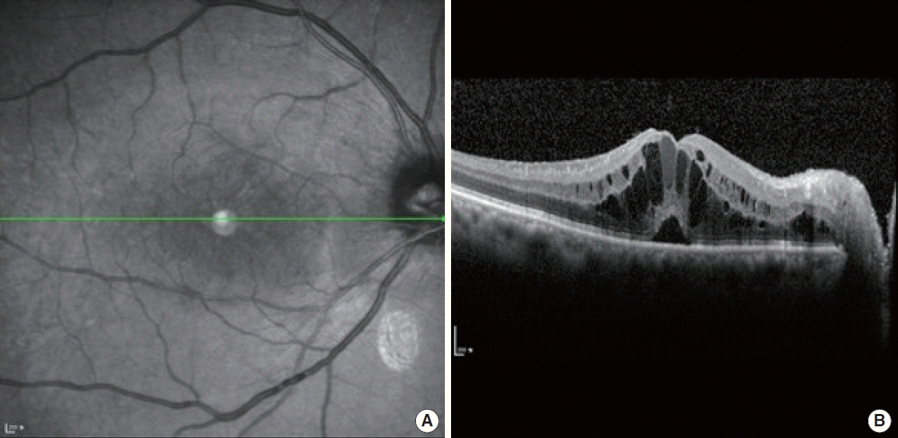
(A, B) The optical coherence tomography image of the right eye 10 months after scleral fixation of intraocular lens (IOL) due to IOL dislocation. Multiple large, hyporeflective cystic spaces are seen within the outer plexiform layer and small cystic cavities are seen within the inner plexiform layer. Also, accompanying subfoveal neurosensory detachment is noticed.
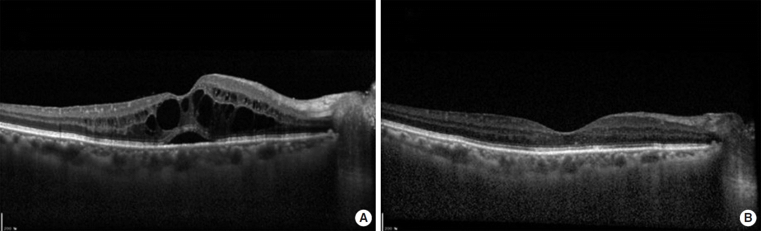
Optical coherence tomography image of recurrent macular edema after intravitreal bevacizumab and triamcinolone injection (A) in the right eye. Significant improvement of macular edema 1 month after intravitreal Ozurdex (Allergan Inc., Irvine, CA, USA) implantation (B).

Anterior segment photograph showing dexamethasone implant (Ozurdex; Allergan Inc., Irvine, CA, USA) in the anterior chamber with clear cornea without edema.
고 찰
덱사메타손 임플란트는 유리체강 내 삽입하여 덱사메타손을 지속적으로 방출하면서 생분해되는 물질로 망막정맥폐쇄와 동반된 황반부종의 치료에 있어 유의한 치료효과가 입증되어 최근 많이 이용되고 있는 치료법이다[1]. 백내장 수술 후 수정체 후낭 파열이 동반되어 있거나 수정체소대의 손상이 있는 경우에 유리체강 내로 주입한 임플란트가 전방 내로 이동할 수 있고 만약 유리체절제술이 되어있는 눈이라면 완충작용을 해 줄 유리체가 없어 임플란트가 전방 내로 더 쉽게 이동할 수 있다. 고농도의 덱사메타손은 세포 괴사와 세포 자살에 영향을 미쳐 각막내피세포에 손상을 주는 것으로 알려져 있는데[15], 전방으로 이동한 덱사메타손 임플란트도 각막내피에 직접 닿거나 높은 약물 농도로 인해 각막내피세포를 손상시켜 각막부종을 일으킬 수 있을 것이다. Ozurdex의 약물 설명서를 보면 수정체 후낭의 손상이 있을 경우 임플란트가 전방으로 이동할 수 있다는 주의사항이 적혀 있고 안구나 안구 주위 염증이 있는 경우, 진행된 녹내장이 있는 경우, 수정체 후낭 파열이 동반된 무수정체안, 수정체 후낭 파열이 동반된 전방인공수정체안 그리고 과민반응이 있는 환자에서는 유리체강 내 주입술이 금기로 명시되어 있다.
Khurana 등[8]의 보고에 의하면 덱사메타손 임플란트의 전방탈출을 보인 15명 18안에서 주입술을 시행하고 평균 13일(짧게는 5일, 길게는 44일) 뒤에 전방탈출이 관찰되었고, 그중 16안에서 각막부종이 관찰되었다. 그중 10명의 환자(71%)에서 임플란트의 제거에도 불구하고 각막부종이 호전되지 않았고 6명의 환자(43%)는 각막이식을 필요로 하게 되었다. Pardo-López 등[9]은 백내장 수술 중 수정체 후낭이 파열되어 전유리체절제술을 시행하고 홍채 고정 인공수정체를 삽입한 눈에서 분지망막정맥폐쇄가 발생하여 유리체강 내 덱사메타손 임플란트 주입술을 시행하고 3주 뒤 임플란트가 전방 내로 이동하여 심각한 각막부종과 각막부전을 가져와 결국 각막이식을 하였다고 보고했으며, Bansal 등[10]의 보고에서도 유리체절제술과 수정체제거술을 시행한 무수정체안 3안에서 임플란트의 전방탈출로 1안에서는 심각한 각막부종이 발생하여 임플란트의 제거를 필요로 하였다.
본 증례에서는 수정체 후낭 결손이 있는 환자에서 임플란트를 주입하고 70일이 지나서 임플란트의 전방탈출이 관찰되었는데, 각막부종이 관찰되지 않고 약물의 치료효과는 유지되고 있어서 제거를 시도하지 않고 안압 조절을 하면서 경과관찰을 하였다. 각막 부종이 보이지 않은 것은 덱사메타손 임플란트가 최대 효과를 보이는 60일 이상이 지나 약물효과가 점차 감소되는 시점에 전방으로 이동하여 각막내피에 손상이 크지 않았기 때문일 것으로 생각된다. 본 환자의 각막내피세포검사에서 유리체강 내 덱사메타손 임플란트가 전방으로 탈출하기 전에는 각막내피세포밀도는 1,741개/mm2를 보였고 임플란트가 완전히 흡수되고 난 후 각막내피세포밀도는 1,733개/mm2 정도로 차이를 보이지 않았다(Fig. 4). 만약 주입술 후 1개월 이내에 전방탈출이 발생하고 각막부종이 보인다면 즉시 임플란트를 유리체강 내로 재위치시키거나 제거하는 조치가 필요할 것이다. Vela 등[11]은 30게이지 바늘을 이용해 임플란트를 홍채 뒤로 밀어 넣어주는 방법을 제시하였고 Collet [12]은 수술적 처지 없이 환자의 눈을 산동시켜 머리를 뒤로 젖힌 뒤 손가락으로 안구를 가볍게 두드려 임플란트를 유리체강 내로 재위치시키는 방법을 제시하였다.

Specular microscopy before the second migration of Ozurdex (Allergan Inc., Irvine, CA, USA) into the anterior chamber (A) and after complete resolution of the implant (B).
수정체 후낭의 결손으로 인공수정체 공막고정술과 유리체절제술을 이전에 시행 받은 환자에서 유리체강 내 덱사메타손 임플란트 주입술을 시행하면 임플란트의 전방탈출이 발생할 수 있으므로 좀 더 자주 관찰할 필요가 있다. 또한 덱사메타손 임플란트 주입술 후 60일 이상 경과하여 임플란트가 전방으로 탈출하였을 경우에 스테로이드 치료효과를 위해 임플란트를 제거하지 않고 경과를 지켜볼 수 있을지에 대해서는 향후 추가적인 연구가 필요할 것이다.
감사의 글
이 연구는 순천향대학교 연구비 지원을 받아 수행되었다.