An Inguinal Endometriosis without Any Other Pelvic Endometriosis Mimicking Direct Inguinal Hernia: A Case Report
Article information
Abstract
Inguinal endometriosis is a rare disease. Patients with inguinal endometriosis usually exhibit cyclic inguinal pain with a cyclic change in the size of the inguinal mass. It is more often found on the right side and commonly accompanies concomitant endometriosis lesions on pelvic organs or peritoneum. We report a case of inguinal endometriosis without any of the usual characteristics. A 48-year-old with non-severe dysmenorrhea and pelvic pain presented a left inguinal mass, palpable only when standing. Under the impression of direct inguinal hernia, laparoscopic herniorrhaphy was performed. Intraoperative laparoscopic findings revealed no other endometriosis lesion on the pelvic organ or in the abdominal cavity, and the histopathologic report confirmed it was endometriosis. Thorough inspection and excision of endometriosis lesions in the pelvic cavity are crucial for treatment. Therefore, an appropriate surgical plan following accurate preoperative diagnosis is important. If the intraoperative evaluation of endometriosis was not enough, postoperative gynecologic assessment is strongly recommended.
INTRODUCTION
Endometriosis is a disease when endometrial glands and stroma are presented outside the normal endometrial cavity and musculature [1]. Dr. Allen in 1896 first described endometriosis presenting as a groin lump [2]. Inguinal endometriosis is rare, counting up only 0.6% of endometriosis patients [3]. Patients with inguinal endometriosis usually complain of palpable inguinal mass that is usually associated with cyclic pain and cyclic change in size [4].
We would like to report a case of inguinal endometriosis of a 48-year-old female patient mimicking an inguinal hernia, without cyclic inguinal pain or change in size. Based on the clinical characteristics and that there were no other endometriosis lesions on pelvic organs and peritoneum, it was hard to suspect the inguinal mass to be endometriosis.
CASE REPORT
A 48-year-old woman visited the surgery clinic presenting a palpable left inguinal mass. The patient noticed it 10 days ago and it was painless. Obstetric history includes two vaginal deliveries. The patient has experienced dysmenorrhea which subsided with oral non-steroidal anti-inflammatory drugs, and non-severe chronic pelvic pain irrelevant to the menstrual cycle.
The patient went under right ovarian cystectomy with a lower midline incision 26 years ago, which was diagnosed as mature cystic teratoma, and received reoperation 2 days after due to a hematoma formation within the incision.
The patient stated the inguinal mass was felt to the size of a ping-pong ball and it was soft at first, but got harder over time. The patient could only feel the mass while standing upright, not when lying down. The mass was palpable on the left inguinal area on the physical exam, and it bulged out with the Valsalva maneuver. It was easily manually reduced.
Ultrasonogram (USG) was performed with the impression of an inguinal hernia. The USG exam revealed a 3-cm-sized solid mass with a cystic portion is noted in the left suprapubic subcutaneous layer (Fig. 1). Abdominopelvic computed tomography (APCT) showed a 3-cm-sized enhancing solid lesion noted in the left inguinal region contiguous to the left pelvic cavity without fluid collection (Fig. 2A–D). Transvaginal USG revealed no abnormal findings in both ovaries.
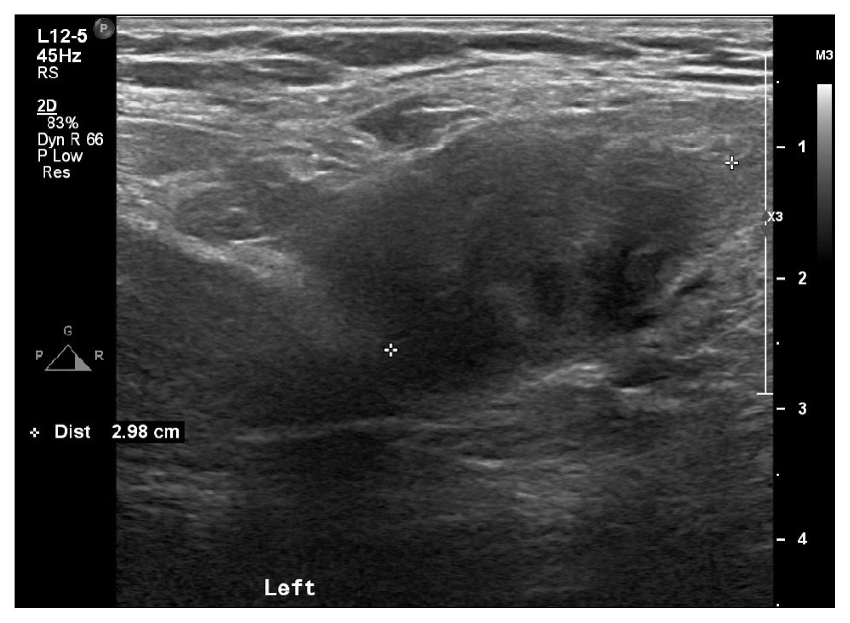
Ultrasonogram image of the inguinal mass. A unilocular mass with heterogeneous ground glass echogenicity of the cyst content (30×18 mm) is noticed in the left suprapubic subcutaneous layer.

Abdominopelvic computed tomography images. (A) Coronal, (B) sagittal, and (C) axial scan shows ovoid left inguinal mass (41×21 mm) with irregular enhancement with central hypoattenuation (dotted white circle). (D) Axial scan shows the inguinal mass is connected to the left round ligament (white arrow).
The surgery department started the diagnostic laparoscopy. Adhesions between the sigmoid colon and the left paracolic gutter were noticed and adhesiolysis was done. After pulling down the left round ligament of the uterus, a firm 3×2 cm-sized myomatous-looking mass was retracted from the left inguinal canal (Fig. 3A–C). The gynecology department joined the surgery and did a laparoscopic inspection of other pelvic organs and the peritoneum. No other endometriosis lesion was noticed.

Intraoperative images. (A) After the adhesiolysis of sigmoid colon, the thickened and tightened left round ligament of the uterus (UT) was noted (white arrowheads). (B) Pulling down the left round ligament, the mass dragged down from the inguinal canal (white arrows). (C) A bulge at the internal inguinal ring after resecting the mass and the extraperitoneal portion of the left round ligament (white circle).
After en bloc resection of the mass, we noticed the peritoneal outward bulge like a direct inguinal hernia, so we performed herniorrhaphy with a polypropylene mesh. A histopathologic exam revealed the mass was endometriosis (Fig. 4A–C). After the surgery, there was a no more palpable mass on the left groin. The patient was not in a post- or peri-menopausal state at the time, so we prescribed dienogest 2 mg daily for 3 months to prevent the recurrence of endometriosis and to reduce chronic pelvic pain and menstrual pain. After 3 months, the decision was made to stop the medication due to the side effects of abnormal uterine bleeding. There is no evidence of recurrence so far for 3 years.

Histopathologic findings. (A) The macroscopic finding of the specimen. (B) Hematoxylin and eosin (H&E)-stained section of excised tissue showing typical endometrial glands and stroma (×100). (C) H&E stain of the same finding (×200).
The patient had provided informed written consent for publication of the case.
DISCUSSION
Patient with inguinal endometriosis often exhibits a history of previous gynecologic or obstetric surgery, and usually are multiparous women [5]. The patient of this case also experienced open ovarian cystectomy and additional surgical hematoma removal 2 days 26 years ago. People with no operation history, however, are also reported to have inguinal endometriosis [6].
This case can be considered rare among inguinal endometriosis cases since the lesion was found in the left inguinal canal and there was no distinctive endometriosis lesion on any other pelvic organs or pelvic peritoneum.
Inguinal endometriosis is more commonly found on the right side. The reason for this is thought to be the presence of the sigmoid colon which works as a preventive force on the left inguinal area [5]. Based on the literature review of Alsinan et al. [6], inguinal endometriosis is found less often on the left side, which was 13 out of 81 cases.
In the most cases of inguinal canal endometriosis, concomitant intraperitoneal or pelvic endometriosis is reported [2]. Pre-existing pelvic endometriosis may have had an effect on the round ligament to be a route of inguinal endometriosis. Other possible origins, especially when the round ligament endometriosis is absent, are thought to be herniation of intra-abdominal endometriosis in the hernia sac [7].
Preoperative differential diagnosis is important because selecting an appropriate surgical approach is necessary. Possible differential diagnosis is inguinal hernia; hydrocele of the canal of Nuck; lymphadenopathy; and tumors such as lipoma, neuroma, lymphoma, and cancer [8].
APCT is a strong modality not only to confirm the diagnosis of inguinal endometriosis but also to exclude other possible diagnoses [9]. Still, magnetic resonance imaging is thought to be the most specific and sensitive tool for the diagnosis of endometriosis since it can detect iron particles in the hemosiderin within endometrioma [4]. On the APCT of this case taken preoperatively, the first impression was endometriosis of the left inguinal lesion.
To treat inguinal endometriosis, complete surgical resection of the mass and the extraperitoneal portion of the round ligament is essential because if even a portion of the lesion is left behind, residual symptoms and recurrence can occur [10]. The extraperitoneal portion of the round ligament is easily affected by endometriosis and rarely detected [3]. We made sure to excise the whole mass with the extraperitoneal portion of the round ligament which caused wide defect after excision so that hernia repair with a mesh was necessary (Fig. 4C).
Since inguinal endometriosis has a combined nature of inguinal hernia and endometriosis, the two departments of surgery and gynecology are encouraged to be involved together in the surgical treatment. If the thorough laparoscopic inspection of the pelvic organs and peritoneum by a gynecologist was not possible at the time of operation, it is recommended to refer the patient for gynecological assessment postoperatively [7].
The necessity of postoperative hormonal therapy when there was only inguinal endometriosis without any pelvic endometriosis is controversial. It is sometimes recommended as an adjuvant therapy to lower the risk of recurrence [7]. In this case, we decided to prescribe the daily dienogest 2 mg to ease the chronic pelvic pain and dysmenorrhea, even though the patient did not have other pelvic endometriosis.
ACKNOWLEDGMENTS
The doctor Woo-seok Kim (Department of Surgery, Soonchunhyang University Gumi Hospital, Gumi, Korea) participated in patient care (the surgery doctor who did herniorrhaphy) and had not contributed to the writing of the article.
Notes
No potential conflict of interest relevant to this article was reported.