경골 염전과 슬관절 내반각: 무지외반증의 병인인가?
Tibial Torsion and Knee Varus Angle: Are These Aetiologies of Hallux Valgus?
Article information
Trans Abstract
Objective
The purpose of this study is to evaluate that knee deformity (varus or valgus) due to osteoarthritis of the knee and tibial torsion could be aetiologies of hallux valgus.
Methods
Forty-nine patients (43 females, six males; mean patient age, 69.88 ± 6.12 years) before total knee arthroplasty for advanced primary osteoarthritis were recruited. All deformities were of the left knee. Preoperative torsional computed tomography, anteroposterior (AP) and lateral weight-bearing foot radiographs, AP and lateral weight-bearing knee radiographs, and long-leg standing AP views were obtained for each patient. The correlations between foot angle and knee varus angle or tibial torsion angle were examined.
Results
There was no significant correlation between knee varus angle and foot angle. Similarly, there was no significant relationship between tibial torsional angle and foot angle, except talocalcaneal angle (TCA)-lateral (r=0.28). No significant relationships were found between TCA-AP and (1–2 intermetatarsal angle [IMA] and hallux valgus angle [HVA]), or between TCA-lateral and (1–2 IMA and HVA).
Conclusion
No significant correlations were found between the knee anatomical axis (knee varus angle) and foot angle or between the tibial torsional angle and foot angle. Tibial torsion and knee varus angle were not aetiologies for hallux valgus.
서 론
무지외반증은 성인에게 나타나는 퇴행성 장애로 생각되고 있다[1]. 무지외반증이 생기는 병인으로 신발[2], 직업[3], 유전[3-5], 평발[6], 중족-설상골 관절의 과운동성[7,8], 인대 이완[4,9], 아킬레스건 구축과 몸무게가 있다[10]. 하지만 모든 병인이 완전하게 밝혀지지 않았다. 경골의 염전은 하지에서 정강이의 축면이나 횡단면 정렬을 나타낸다. 과도한 외측 경골 염전은 족부의 외측 각도와 연관될 수 있으며, 이는 성인에서 외반 편평첨족으로 인한 부정렬을 부분적으로 진행하게 하거나, 무지외반 부정렬, 중족골 퇴행성 관절염을 진행하게 할 수 있다[11-14]. 그러나 아직 경골 염전과 무지외반 변형의 관계에 대해 연구된 논문은 없다. Ledoux와 Hillstrom [15]은 무지외반증과 동측 슬관절의 내측 골관절염의 연관성에 대해 보고하였는데, 이는 내측으로 전위된 지면 반발력이 슬관절 내측의 모멘트 암을 증가하여 발생되는 것으로 발표하였다. 골관절염으로 인한 슬관절의 내반, 외반 변형이 무지외반증과 함께 고려된 적이 없으며 병인으로 밝혀진 적이 없다. 이 논문의 목적은 골관절염으로 인한 슬관절의 내반, 외반 변형과 경골의 염전이 무지외반증의 병인이 될 수 있는지 밝히는 것이다. 우리는 슬관절 부정렬과 경골 염전이 무지외반 변형에 영향을 미친다고 가정하였다.
대상 및 방법
순천향대학교 의과대학 부천병원의 임상연구심의위원회의 승인을 얻은 후(SCHBC_IRB_2010-42), 2011년 1월부터 6월까지 진행된 원발성 골관절염 환자를 대상으로 슬관절 전 치환술을 시행하기 전에 52명의 환자를 대상으로 연구를 진행하였다. 류마티스관절염이나 이차성 골관절염과 같은 다른 진단을 받은 환자를 제외하였고, 임상 연구자가 환자의 임상정보를 수집했다. 52명의 환자 중 3명이 제외되었는데, 1명은 컴퓨터단층촬영(computed tomography, CT)을 거부하였고, 다른 1명은 체중부하 방사선사진을 찍을 수 없었으며, 마지막 1명은 참여를 거부했다. 결과적으로 49명의 환자가 최종 분석에 포함되었다. 모두 43명의 여성과 6명의 남성 환자가 포함되었으며, 환자의 평균 연령은 69.88세(표준편차[standard deviation]=6.12)였다. 모든 기형은 왼쪽에 있었다.
모든 환자는 슬관절 전치환술을 위해 입원 시 동일한 수술 전 평가 프로토콜을 사용하여 검사하였다. 수술 전 검사로는 염전 CT, 전후방 및 측방 체중부하 족부 방사선사진, 전후방 및 측방 체중부하 무릎 방사선사진, 그리고 체중부하 하지 전장 전후방 방사선사진이 포함되었다.
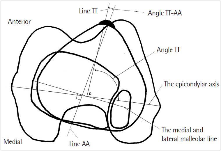
염전 CT는 Nagamine 등[16]의 방법으로 시행하였다. 환자는 CT 기계에서 앙와위로 위치한 뒤 하체는 경골의 장축이 침대와 시상면과 관상면으로 평행하도록 하였다(Fig. 1). 슬개골 인대 부착부위의 경골결절과 발목관절에서 관상면으로 1 cm 근위부에서 스캔을 시행하였다. 경골결절 내측 1/3 지점과 과상간선의 중앙을 이은 선(line TT)을 지정하였고, 두 번째 선은 발목관절에서 1 cm 근위부에서 내과와 외과의 중앙점을 이은 선(line AA)을 지정하였다. 두 개의 선을 얻은 절단면을 겹쳐서 line TT와 line AA의 각(angle TT-AA)을 측정하였다(Fig. 2). 체중부하 하지 전장 전후방 방사선사진에서 대퇴골과 경골의 종축 사이의 해부학적 축과 슬관절 내반 각도를 측정하였다. 족부 방사선사진은 환자의 체중부하상태로 시행하였다.

The subject was placed supine on the computed tomography scanner, and the lower extremity was positioned so that the long axis of the tibia was parallel to the bed in the sagittal and coronal planes with 30˚ knee flexion. Written informed consent was obtained from the patient.

Two images of computed tomography scans were superimposed and angle TT-AA was measured. Line TT was the line from the medial 1/3 of the tibial tuberosity to the center of the epicondylar line. Line AA was the line perpendicular to the medial and lateral malleolar lines. Angle TT-AA was the angle between line TT and line AA.
체중부하 전후방 방사선사진에서 첫 번째 중족골과 근위 지골의 축은 골의 내측 및 외측 피질에서 등거리인 골간부위를 이등분하는 선을 축으로 구하고, 이 축들의 교차에 의해 생성된 각도를 무지외반각(hallux valgus angle, HVA)이라 하였다[17]. 동일한 방식으로 제1, 2 중족골간각(1–2 intermetatarsal angle, IMA)을 구하였다. 또한 거골과 종골에 적용하여 전후방 거종골간각(talocalcaneal angle-anteroposterior, TCA-AP)을 구하였다.
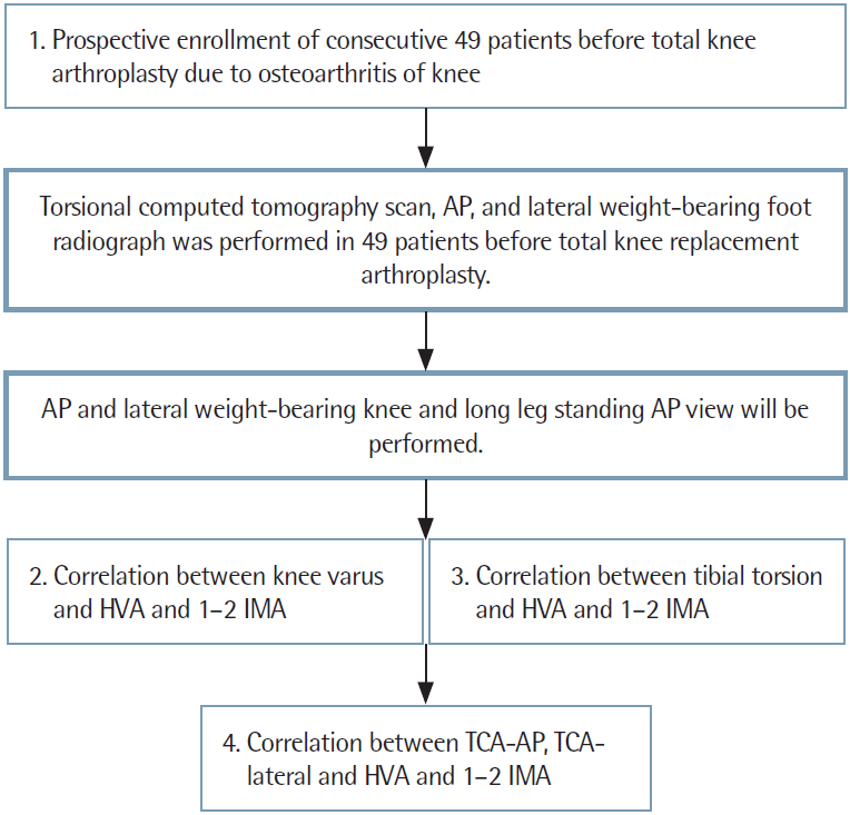
체중부하 측방 방사선사진에서 거골과 종골의 내측 및 외측 피질에서 등거리인 골간부위를 이등분하는 선을 축으로 구하고, 이 축들의 교차에 의해 생성된 각도를 외측 거종골간각(TCA-lateral)이라 하였다. 족부 체중부하 방사선사진으로 얻어진 수치를 슬관절 내반각, angle TT-AA와의 상관관계에 대해 확인하였다(Fig. 3).

Flow chart of the knee varus angle and tibial torsion-hallux valgus. AP, anteroposterior; HVA, hallux valgus angle; IMA, intermetatarsal angle; TCA, talocalcaneal angle.
데이터 관리 및 통계분석은 SPSS ver. 12.0 (SPSS Inc., Chicago, IL, USA)을 사용하여 수행되었다. 모든 값은 연속변수의 경우 평균±표준편차로, 범주변수의 경우 피험자 수(%)로 표시하였다. Angle TT-AA와 1–2 IMA, TCA-AP, TCA-lateral 및 HVA 사이의 선형 상관관계를 설명하기 위해 피어슨 상관계수를 얻었다. 또한 HVA와 각의 선형 상관관계는 피어슨 상관분석을 사용하여 평가하였다. 정상 및 비정상군 간의 TT-AA 값의 차이는 카이제곱검정 또는 피셔의 정확검정을 사용하여 분석하였다. 0.05 미만의 P값을 통계적 유의성의 최소수준으로 취했다. 모든 가설검정은 양면검정이었다.
HVA와 IMA 간의 상관관계를 평가할 수 있는 충분한 전력을 확보하기 위해 PASS 2008 (NCSS, Kaysville, UT, USA) 소프트웨어를 사용하여 피어슨 상관계수 값을 기반으로 한 전력분석을 수행하였다. 귀무가설 상관계수가 0이고 유의수준이 0.05인 것으로 가정하면, 대안적 가설하에서 피어슨 상관계수가 0.664 이상인 경우 15명의 피험자의 표본크기가 80%의 기회를 얻었다.
결 과
슬관절 내반각과 족부각의 상관분석 결과 통계적으로 유의하지 않은 열등한 상관관계를 보였다(Table 1). 평균 슬관절 내반각은 0.84˚(최대=10.5˚, 최소=-9.2˚), 평균 TCA-AP는 29˚(최대=55.4˚, 최소=8.9˚)였고, 평균 TCA-lateral은 23.9˚(최대=40.7˚, 최소=17.7˚), 평균 HVA는 16.6˚(최대=52.8˚, 최소= 0.6˚), 평균 1–2 IMA는 9.4˚(최대=16.9˚, 최소=3.8˚)였다.
경골의 염전각과 족부각의 상관관계 분석에서도 TCA-lateral (r= 0.28)을 제외하고는 통계적으로 유의하지 않은 열등한 상관관계가 발견되었다(Table 2). 평균 경골 염전각은 6.4˚(최대=20.42˚, 최소=-18.84˚)였다.
TCA-AP와 1–2 IMA 및 HVA 간의 상관관계 분석은 통계적으로 유의하지 않은 열악한 상관관계를 발견했다. TCA-lateral과 1–2 IMA 및 HVA도 유사한 결과가 나타났다(Table 3).
고 찰
경골 염전은 하지에서 정강이 부위의 축 방향 또는 횡 방향 평면정렬을 나타낸다. 태아에서 재태 4주차에 하지 싹이 형성되기 시작한다[18,19]. 재태 7주차에 하지는 내측으로 회전하며 무지가 정중선으로 위치하게 된다. 그 후 외측과 외회전이 서서히 일어나며 골성숙이 일어난다. 경골 염전은 출생 시 약 5˚가량 외회전되어 있고, 골격이 성숙되며 15˚가량 외회전되게 된다[20]. 과도한 경골의 내회전상태는 보행 시 족부의 충격흡수 기전을 방해하여 관절에 충격을 줄 수 있다. 또한 과도한 경골 내측 염전은 족부의 내측 각을 악화시켜 성인에서 슬관절 내측 골관절염과 연관될 수 있다[11,21]. 과도한 경골 외측 염전은 족부의 외측 각을 악화시켜 성인에서 부분적으로 외반 편평첨족으로 부정렬, 무지외반 부정렬과 중족 관절의 퇴행성 골관절염을 초래할 수 있다[11-14]. 경골의 내측 염전은 경골 말단의 골간단 회전에 의해 유발된다. 발은 일본식 좌식으로 내회전되며[16], 한국인의 생활양식도 일본과 유사하다. 한국인도 바닥에 똑바로 앉는 생활을 한다. 이 연구에서 평균 경골 염전 각은 6.4˚(최대=20.42˚, 최소=-18.84˚)로 경골의 내측 염전을 나타내었고, 이는 Nagamine 등[16]의 결과와 유사하다. 과도한 경골 외측 염전은 족부의 외측 각을 악화시켜 성인에서 부분적으로 외반 편평첨족 부정렬에 영향을 줄 수 있다[11-14]. 그러나 본 연구에서 경골의 내측 염전과 발 각과의 상관관계는 유의하지 않았다. 과도한 경골 외측 염전은 외반 편평첨족 부정렬과 무지외반 부정렬에 영향을 미치지만, 동양인은 보통 생활방식으로 인해 경골 내측 염전이 발생한다[16]. 따라서 동양인의 경골 염전은 보통 발의 변화와 무지외반 부정렬에 영향을 주지 않는다
슬관절 골관절염은 생화학적 및 기계적 악화에 의해 유발될 수 있는 파괴 및 복구 기전을 모두 포함하는 대사활성의 역동적인 질병이다. 확인된 생체역학적 변화는 관절과 연부조직의 퇴행성 변화에 대한 반응, 또는 질병과정에서 보상 메커니즘을 포함한 질병의 발달로 인한 것으로 보인다[22,23]. 체위성 외반 편평족은 무릎의 기계적인 과부하에 대한 감수성을 증가시켜 슬개골 대퇴관절 통증을 발생시킨다[24]. 이것은 슬관절의 내측으로 모멘트 암이 증가하여 지면 반발력을 내측이 받기 때문에 무지외반증과 슬관절 내측의 골관절염의 연관을 시사하고 있다[15]. 본 논문의 결과에서 평균 슬관절 외반각은 0.84˚(최대=10.5˚, 최소=-9.2˚)였고, 이는 다양한 하지 각도를 가지고 있으며 심한 슬관절 내반 변형이 없었다. 이 결과를 바탕으로 우리는 무지외반증이 슬관절 골관절염에 영향을 미칠 수 있지만 슬관절의 해부학적 축(무릎 내반각)은 족부각에 영향을 미치지 않는다고 가정했다. 무지외반증은 첫 번째 중족골의 내측 편위와 엄지 발의 측방 편위가 있는 첫 번째 중족 지간 관절의 정적 아탈구이다. 따라서 골관절염에 의한 슬관절 내과 각도는 역동적인 질환으로 발의 각도에 영향을 미치지 않는 것으로 보인다.
편평족과 무지외반증과의 연관성은 논란의 여지가 있다[1,6,25-27]. Tanaka 등[6]이 편평족과 무지외반증과의 연관성에 대해 보고하였지만, 다수의 저자가 무지외반증 환자에서 편평족이 정상군보다 적을 수 있음을 언급하였다[1,25-27]. 우리는 경골 염전과 편평족 사이의 상관관계가 많은 연구에서 통계적으로 유의할 것으로 가정하고[11-14], Tanaka 등[6]이 발표한 편평족과 무지외반증의 연관성에서 무지외반증과 경골 염전이 상관관계가 있을 것으로 가정하였으나, 통계학적으로 연관관계를 찾을 수 없었다. 따라서 경골염전과 슬관절 내반각이 무지외반증의 병인학적 요인이 아님을 알 수 있었다.
본 논문의 제한점으로는, 첫 번째로 발목의 축은 슬관절의 굴곡-신전에 영향을 받아서 90˚ 굴곡 시 20˚ 외회전, 슬관절 완전 신전 시 40˚ 외회전을 보이게 되는데[28], 연구계획상 30˚ 굴곡하여 CT를 시행하였기 때문에 슬관절의 회전 정렬에 대해서는 고려되지 못하고 경골 염전에 대해서만 확인하게 되었으며, 두 번째는 2명의 연구자가 모든 각도를 측정하고 평균 각도를 기록했지만, 관찰자 간 및 관찰자 편향이 있을 수 있다는 점이다. 마지막으로 동양인만 조사하였기 때문에 대개 경골의 외회전을 가지는 서양인에 대해 조사할 수 없었다. 추가적인 서양인에 대한 조사가 필요할 것으로 보인다.
결론적으로, 슬관절 해부학적 축(슬관절 내과 각도)과 족부각간의 상관관계는 유의하지 않았으며, 과도한 경골 외측 염전은 외반첨족 변화와 무지외반 부정렬에 영향을 미치지만, 동양인은 생활방식으로 인해 경골의 내측 염전이 발생하므로 동양인의 경골 염전은 보통 발의 변화와 무지외반 부정렬에 영향을 주지 않는다. 경골염전각과 족부각 사이에는 유의한 상관관계가 없었다. 무지외반증은 첫 번째 중족골의 엄지 발의 측부 편위와 내측 편위를 가진 첫 번째 중족 지간 관절의 정적 아탈구이다. 따라서 골관절염에 의한 무릎 내과 각도는 족부각에 영향을 줄 수 없다.


