Estimation of Cardiac Index: Validation of the Mobil-O-Graph NG in Comparison with the FloTrac/Vigileo
Article information
Abstract
Objective
Although the reference value of cardiac index (CI) is derived by pulmonary arterial pressure, the use of pulmonary arterial catheterization is limited by low cost effectiveness and many concerns regarding complications. Therefore, relatively noninvasive indirect measurement is used widely perioperatively. The goal of this study was to determine the accuracy of the CI derived by Mobil-O-Graph NG (cCI) noninvasively in patients undergoing general anesthesia by comparing that measured by FloTrac/Vigileo (fCI), the minimal invasive method.
Methods
The Bland–Altman method was used to quantify agreement. Bias (mean difference between fCI–cCI) represents the systematic error between methods and precision (standard deviation of the bias) represents the random error or variability between techniques. The percentage error was considered clinically acceptable, and the tested method (Mobil-O-Graph NG) was regarded as interchangeable with the reference method (FloTrac/Vigileo), if it was below 30%.
Results
One hundred and ninety-five patients were included in this study, and CI, measured in the 121 patients. The Bland–Altman analysis revealed a bias −0.01 and the percentage error of 32.4%. And the difference is inversely increased according the mean CI.
Conclusion
Results showed that CI measured by Mobil-O-Graph NG had a wide limit of agreement with that measured by FloTrac/Vigileo, therefore regarded as not interchangeable.
INTRODUCTION
Although the reference value of cardiac index (CI) is derived by pulmonary arterial pressure, the use of pulmonary arterial catheterization is limited by low cost effectiveness and many concerns regarding complications. Therefore, relatively noninvasive indirect measurement is used widely perioperatively. The FloTrac/Vigileo (Edwards Lifesciences, Irvine, CA, USA) is a less invasive monitor and measures pulse pressure (PP)-derived CI without external calibration. The accuracy is validated in many studies [1–3]. However, there are also concerns about the accuracy [4–6].
Although mean blood pressure and diastolic blood pressure are relatively constant in the conduit arteries, it has been known that systolic blood pressure (SBP) and PP are higher in the peripheral than in the central arteries. This so-called SBP or PP amplification is the consequence of the progressive reduction of diameter and increase in stiffness from the proximal to the distal arterial vessels and mostly of the modification in the transit of wave reflections. It seems obvious that central pressures are more relevant than peripheral pressures for the pathogenesis of cardiovascular disease: it is central SBP against the heart ejects (afterload), and it is central PP that distends the large elastic arteries [7]. Thus, while being highly correlated, central pressures cannot be reliably inferred from peripheral pressures. Mobil-O-Graph NG, (IEM, Stolberg, Germany) measures central pressures using brachial waveforms and the ARCSolver algorithm and estimates CI. This value derived based on the central blood pressure might have good performance to estimate the CI.
The goal of this study was to determine the accuracy of the CI derived by Mobil-O-Graph NG in patients undergoing general anesthesia by comparing that measured by FloTrac/Vigileo to investigate the interchangeable.
MATERIALS AND METHODS
We included all types of surgery using FloTrac for monitoring if both arms are available for measurement. Exclusion criteria were (1) more than mild mitral regurgitation, (2) more than mild aortic regurgitation, (3) arrhythmias, or (4) <18 years old. All patients gave informed consent to participate in the study.
All the data were obtained while the patient was in a supine position and general anesthesia. And a transducer (TruWave transducer, Edwards Lifesciences) was positioned at the midaxillary line. After an arterial catheter was inserted into the radial artery and attached to the FloTracTM device, arterial pressure waveforms were sent simultaneously to the Vigileo. We used fourth-generation FloTrac/Vigileo and only high-quality recordings of the radial waveforms by visual inspection and by a device-specific quality index 80% were used. Any time during the operation, when we can obtain qualified CI of FloTrac (fCI), we cuffed the opposite arm and start the measurement of Mobil-O-Graph NG. Two size of Mobil-O-Graph NG cuff prepared, and we used proper size for patient. We set the measuring frequency 30 times at one hour and run it for 4–5 minutes. Two iterations were performed and the particular estimates for CI of Mobil-O-Graph NG (cCI) were averaged. The Bland–Altman method was used to quantify agreement. Bias (mean difference between the test method and the reference method: fCI–cCI) represents the systematic error between methods and precision (standard deviation [SD] of the bias) represents the random error or variability between techniques. Limits of agreement were calculated as bias ±2SD and defined the range in which 95% of the differences between methods were expected to lie. The percentage error was calculated as the ratio of 2SD of the bias to the mean CI of the reference method. The percentage error was considered clinically acceptable, and the tested method (Mobil-O-Graph NG) was regarded as interchangeable with the reference method (FloTrac/Vigileo), if it was below 30%, as proposed by Critchley and Critchley [8].
RESULTS
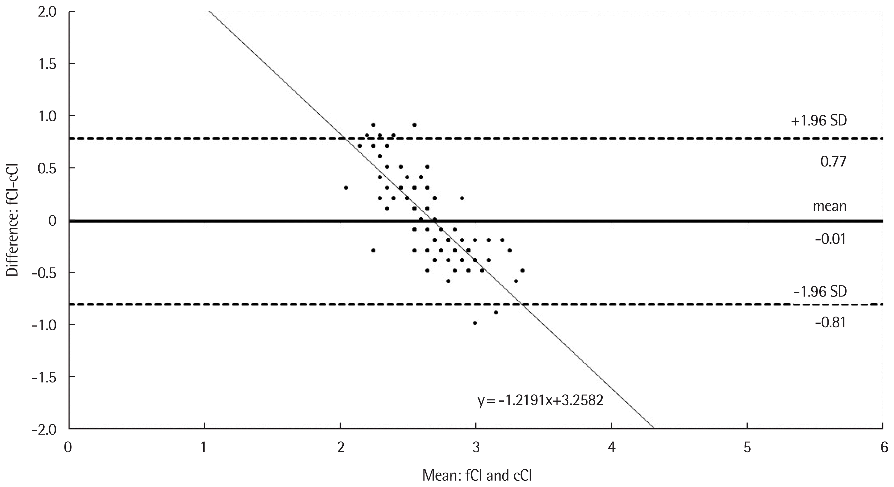
One hundred and ninety-five patients were included in this study, and qualified CI measured in the 121 patients. Fig. 1 shows the Bland–Altman analysis results for comparisons between fCI and cCI for all measures, which revealed a percentage error of 32.4%. And the difference is inversely increased according the mean CI. And the difference is inversely increased according the mean CI.
DISCUSSION
The bias between fCI and cCI are small. However, the Bland–Altman analysis revealed that CI measured by Mobil-O-Graph NG had a wide limit of agreement with that measured by FloTrac/Vigileo, which resulted in a percentage error of 32.4% over the acceptance threshold. And the difference is inversely increased according the mean CI.
The minimally invasive FloTrac/Vigileo system (Edwards Lifesciences) was introduced in 2005 [9]. The device can accurately detect hemodynamic instability associated with changes in systemic vascular resistance [10], especially with the latest fourth generation software [1]. Cardiac output monitoring based on pulse contour analysis (Vigileo-FloTrac) has the potential to be used for goal-directed fluid therapy in the perioperative setting [11]. Therefore, the CI of FloTrac/Vigileo is widely used during general anesthesia. Most arterial waveform analyses are based on the Windkessel theory developed by Langewouters et al. [12], although the principle of measurement is slightly different between the different devices. The FloTrac/Vigileo system is based on the principle that aortic PP (difference between systolic and diastolic pressure) is proportional to stroke volume under stable peripheral vascular resistance (SV=K×PP, where K is a scaling multivariate model proportionate to aortic compliance and vascular tone) [12]. The FloTrac/Vigileo system substitutes the SD of arterial pressure sampled at 100 Hz over the span of 20 seconds for PP, providing the algorithm with 2,000 data points for analysis. Therefore, the FloTrac/Vigileo system is derived from the arterial pressure waveform itself. Although, this is minimally invasive method, still need arterial cannulation. That costs potential adverse effects and some skill and equipment. If there is an interchangeable value that can obtain totally invasively, it will be very useful.
It was demonstrated stronger relations of central versus brachial blood pressure, particularly PP, to carotid artery hypertrophy and extent of atherosclerosis. Left ventricular hypertrophy is more strongly related to systolic pressure than to PP. Furthermore, central pressures are more strongly related than brachial pressures to concentric left ventricular geometry [13]. The prognostic value of central SBP has been established recently. Moreover, an algorithm enabling conventional automated oscillometric blood pressure monitors to assess central systolic pressure could be of value. Central systolic pressure, calculated with a transfer-function like method (ARCSolver algorithm), using waveforms using brachial cuff-based waveform recordings, is suited to provide a realistic estimation of central systolic pressure [14]. Mobil-O-Graph NG, (IEM) measures central pressures using brachial waveforms and the ARCSolver algorithm and estimates CI. If it can estimate the CI in perioperative setting, it will be more valuable than invasive measuring values based on the peripheral pressure. However, in this study we could not validate the accuracy because we compare the CI of Mobil-O-Graph NG with that of FloTrac/Vigileo system.
Although the new fourth-generation FloTrac/Vigileo system after increased vasomotor tone was greatly improved compared with previous versions; however, the discrepancy between CI measured by FloTrac/Vigileo and the reference method was still not clinically acceptable in previous studies [1,15], possibly because the accuracy of FloTrac/Vigileo is greatly influenced by vascular tone. And this is the explanation of our result. In this study, the difference is inversely increased according the mean CI. This may be the result of underestimate of CI by FloTrac/Vigileo. Asamoto et al. [16] found that cardiac output measured by FloTrac/Vigileo tended to underestimate the calculated CIs when the CIs were relatively high.
Estimating the cardiac output constitutes an essential part of the hemodynamic monitoring during general anesthesia, as it provides the basis for therapeutic interventions to ensure adequate tissue perfusion. For that purpose, a pulmonary artery catheter using the thermodilution method has been considered a ‘clinical standard.’ Although because of the risk of various complications, including damage to the cardiac valves and pulmonary artery rupture, we could not evaluate this standard CI to compare.
Results showed that CI measured by Mobil-O-Graph NG had a wide limit of agreement with that measured by FloTrac/Vigileo, therefore regarded as not interchangeable. The discrepancy is possibly because of the algorithm of Mobil-O-Graph NG is not for the status under general anesthesia or not accurate. Second, the CI of FloTrac/Vigileo is greatly influenced by vascular tone, and that may be responsible for the trend of bias.
