Isolated Unilateral Hypoglossal Nerve Palsy Following Transoral Endotracheal Intubation for Endoscopic Sinus Surgery
Article information
Abstract
Hypoglossal nerve palsy is a rare complication of endotracheal intubation. The mechanism of nerve palsy is mainly attributed to stretching or compression of the nerve during airway manipulation. The cuff pressure can also contribute to the occurrence of hypoglossal nerve palsy. Since it is often accompanied by other cranial nerve palsies, meticulous overall cranial nerve examination is necessary. The main treatment is supportive with respiratory monitoring. The prognosis is favorable. Majority of patients achieve nearly full recovery of nerve function. Here, we report a case of unilateral hypoglossal nerve palsy following usual, uneventful endotracheal intubation and review the literature.
INTRODUCTION
Transoral intubation is commonly performed for airway maintenance during general anesthesia. Although rare, unilateral hypoglossal nerve palsy (HNP) is a possible complication associated with intubation. Hypoglossal nerve is the 12th cranial nerve that innervates the extrinsic and intrinsic muscles of the tongue, except for the palatoglossus muscle. Dysarthria, dysphagia, and dyspnea can occur as a result of HNP. It can occur alone or in combination with other cranial nerve palsies [1].
We report a case of solitary HNP following usual, uneventful transoral intubation and review the literature. To the best of our knowledge, this is the first case reported at Soonchunhyang University Bucheon Hospital.
CASE REPORT
A 62-year-old, 153-cm, and 63-kg female was admitted for endoscopic sinus surgery due to chronic rhinosinusitis. Her past medical history consisted of hypertension, diabetes mellitus, and major depressive disorder. She took the usual oral antihypertensive drug (amlodipine 5 mg), and intramuscular glycopyrrolate (0.2 mg) was given as premedication on the morning of surgery.
Anesthetic induction was achieved with intravenous 1% propofol 100 mg and target-controlled infusion (TCI) of remifentanil (3.0 ng/mL). After that, train-of-four (TOF) monitoring using M-NMT module (Datex-Ohmeda Inc., Helsinki, Finland) was performed every 10 seconds for evaluating neuromuscular blockade. Full muscle relaxation (TOF count 0) was induced with intravenous rocuronium 40 mg before intubation. A size 7.0 endotracheal tube (ETT; Mallinckrodt Taperguard, Covidien, Dublin, Ireland) was gently inserted using a curved direct laryngoscope (Macintosh #3). The vocal cords were visualized well (Cormack-Lehane laryngoscopic view grade I) and intubation was not difficult. Cuff inflation was conducted using 4 mL of room air. The cuff pressure was measured by tactile estimation of the pilot balloon [2]. The ETT was inserted through the right side of the oral cavity, but it was fixed on the left side during the operation for ease of surgery. Maintenance of anesthesia was achieved with desflurane, oxygen and medical air (FiO2 0.5, total flow 3 L/min), and infusion of remifentanil (TCI dose 1–3 ng/mL). Nitrous oxide was not used. Ephedrine 10 mg and phenylephrine 100 mcg were injected to treat hypotension after intubation. Otherwise, no significant event was observed. The patient was placed in a semi-sitting position during surgery. Total duration of intubation was 105 minutes.
Two days after surgery, the patient complained of a dull sensation on the right face and difficulty with speech. Movement of the tongue to the right side was restricted (Fig. 1A). No other neurologic abnormality was observed. Brain magnetic resonance imaging revealed no specific brain lesion associated with the patient’s symptom (Fig. 2). Intubation-related HNP was diagnosed. The patient was discharged without any treatment for HNP, except a regular outpatient clinic follow-up. More than 95% recovery was observed on the 59th day after surgery (Fig. 1B).

Tongue movement examination. (A) 2 days after surgery, movement of tongue to right side is restricted. (B) 59 days after surgery, nearly full recovery of tongue movement observed.

Brain magnetic resonance imaging at the onset time of hypoglossal nverve palsy. No evidence of acute infarction. (A) Aixal image, (B) axial image, (C) sagittal image, and (D) coronal image.
DISCUSSION
HNP usually appears as a sign of other medical conditions. Keane [3] reported that although the most common cause of HNP was brain tumor, other conditions such as stroke, trauma, infection, multiple sclerosis, and surgery can also give rise to HNP.
HNP can occur solitarily or in combination with other cranial nerve neuroplegias. Tapia syndrome (co-occurrence of HNP and recurrent laryngeal nerve paralysis) is a variant of HNP [1]. Fortunately, there was no evidence of other cranial nerve palsies in this case.
From an anatomical point of view, the following possible injury mechanisms for the extracranial passage of the hypoglossal nerve have been reported [1]: (1) Excessive retraction of the tongue itself can result in damage to the distal motor fibers that supply the tongue. (2) The hypoglossal nerve lies superficially behind the hyoid bone. Nerve compression can occur at the hyoid bone level. (3) The hypoglossal nerve descends along the most protruding lateral part of the transverse process of C1 (the anterior side) [4]. Hypoglossal nerve stretching can occur in this area (Fig. 3).
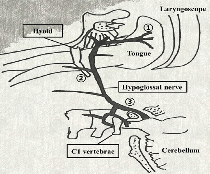
Anatomic considerations for hypoglossal nerve palsy. (1) Excessive retraction of the tongue can result in damage to the distal motor fibers that supply the tongue. (2) The hypoglossal nerve lies superficially behind the hyoid bone. Nerve compression can occur at the hyoid bone level. (3) The hypoglossal nerve descends along the most protruding lateral part of the transverse process of C1. Hypoglossal nerve stretching can occur in this area.
It has been reported that males have a larger hyoid bone and a longer greater cornu of the hyoid than females [1]. Although, our patient was a female, male patients usually have a higher chance of developing hypoglossal nerve damage in the hyoid area due to the above reasons.
Some authors have suggested that high pressure in the ETT or laryngeal mask airway (LMA) can result in hypoglossal nerve injury at the hyoid bone level [1]. When nitrous oxide gas is used, diffusion of nitrous oxide into the ETT cuff can raise the cuff pressure [1]. Nitrous oxide was not used, and we predicted the ETT cuff pressure only by palpation of the pilot balloon in this case. Conventionally, palpation technique has been used widely for the evaluation of cuff pressure because manometers are not always available in daily practice [2]. However, the accuracy of palpation technique depends too much on the operator’s experience and it seems to be unreliable compared to a manometer [2,5]. Kim et al. [6] have reported that more than 1 cm displacement of the ETT is generated by a change in the patient’s head position. Cuff pressure can also change with the head position. Meticulous pressure assessment using a manometer is necessary for surgery that needs head repositioning such as head and neck surgery, shoulder surgery, and spine surgery.
The diagnosis of HNP tends to be delayed. More than half of the patients are diagnosed one day after surgery. Delayed onset of symptoms and residual anesthetic effects may explain this trend [1]. Our patient showed the symptom of HNP two days after surgery. In most cases, airway-related HNP appears to be a self-limiting condition. Shah et al. [1] reported that among the 60 patients, the resolution of symptoms occurred within 6 weeks in 26 patients (43.3%), and within 6 months in an additional 24 patients (40.0%). Some authors have reported that only partial recovery was achieved regardless of the period [1].
Initial supportive care for acute phase HNP consists of respiratory monitoring and oxygen supply [1]. Rehabilitation with speech therapy, exercise training, and electrical stimulation may be helpful [7]. Several authors have emphasized that corticosteroid therapy accelerates recovery from HNP, especially when the airway edema is suspected [1,8]. However, Shah et al. [1] have suggested that corticosteroid therapy does not reduce the follow-up period. In this case, the patient did not present the symptom of airway edema. Therefore, our otolaryngologist decided not to use steroid therapy and just waited for spontaneous recovery with regular outpatient clinic follow-up. The patient achieved more than 95% recovery of the hypoglossal motor function after 59 days of surgery in spite of no specific treatment.
According to the literature, several causes of HNP are suspected. First, we manipulated the laryngoscope with the left hand, and pressed the tongue from the right side to the left side of the oral cavity before intubation. The pressure exerted by this procedure caused stretching of the right side branches of the hypoglossal nerve. It is thought to be the most likely mechanism of HNP in our case. Second, the possibility of the high cuff pressure induced nerve damage was not excluded because we did not use the manometer for evaluating the cuff pressure. Third, compression of the nerve by stretching of ETT during surgery could be suspicious because there was a close proximity between surgical field and ETT insertion site. However, we fixed the tube on the left side of the mouth and the palsy occurred in the right side branch of the nerve. For such reasons, this mechanism could be excluded.
In conclusion, HNP is rare, but it can occur even after usual uneventful orotracheal intubation or LMA insertion. Anesthesiologists should pay careful attention to avoid excessive pressure during all procedures using airway instruments. The depth of ETT and adequacy of cuff pressure should be checked periodically, especially when the patient’s position has changed or surgery time has become longer.