A Case of Pheochromocytoma Initially Manifesting as Acute Myocardial Infarction
Article information
Abstract
Pheochromocytomas are rare catecholamine-secreting neuroendocrine tumors arising from chromaffin cells in the adrenal medulla. Typical classic triad are consisted of headaches, palpitations, and profuse diaphoresis. But some patients with pheochromocytomas have other cardiovascular manifestations such as left ventricular hypertrophy, congestive heart failure, and cardiac arrhythmia. Rarely, pheochromocytomas manifest as acute myocardial infarction leading to delayed diagnosis and treatment. We experienced one case of pheochromocytoma initially manifesting as acute myocardial infarction which showed normal coronary artery on coronary angiography. Pheochromocytoma should be suspected and evaluated in patients with acute myocardial infarction whose coronary angiography shows normal coronary without definite thrombosis.
INTRODUCTION
Pheochromocytoma is neuroendocrine tumor releasing cathecholamines such as norepinephrine, epinephrine, and dopamine and being one of the etiologies of secondary hypertension. Typical triad of symptoms is consisted of headache, diaphoresis, and palpitation. However, persistent or paroxysmal hypertension is the only feature of pheochromocytoma instead of these typical symptoms in many patients. In addition to hypertension, other cardiovascular manifestations such as left ventricular hypertrophy, congestive heart failure, and cardiac arrhythmia are observed in patients with pheochromocytoma. Rarely, pheochromocytoma manifests as acute myocardial infarction leading to delayed diagnosis and treatment. When patients with pheochromocytoma who present this rare cardiovascular manifestation without typical symptoms visit the emergency room, doctors can miss the hidden pheochromocytoma and let the patient’s prognosis get worse leading to being diagnosed at autopsy. We experienced one case of pheochromocytoma initially manifesting as acute myocardial infarction which showed normal coronary artery on coronary angiography. So early diagnosis is very important for appropriate treatment and prognosis of the patients.
CASE REPORT
A 49-year-old woman visited emergency room complaining of retrosternal pain who was transferred from local hospital with the impression of acute coronary syndrome. On past history, she had been taking medicine for type 2 diabetes. At admission, her blood pressure was 90/60 mm Hg and his pulse rate was 102 per minutes. On laboratory findings, each creatine kinase-myocardial band (CK-MB) and troponin-I were elevated by 55.0 ng/mL and 0.065 ng/mL. After follow-up, CK-MB was slightly decreased by 48.3 ng/mL but troponin-I was increased by 0.078 ng/mL. There were about 2 mm ST segment depression with T-wave inversion on precordial leads and inferior leads on electrocardiography at admission day (Fig. 1A). On echocardiography, ejection fraction was decreased by 40% and left ventricular hypertrophy with wall motion abnormality was noted (Fig. 1B). Moreover, follow-up blood pressure was decreased to 70/40 mm Hg. As persistent retrosternal pain, increased cardiac enzymes, and left ventricular dysfunction suggested high risk acute myocardial infarction, emergent coronary angiography was performed and showed normal coronary arteries. Though patient’s clinical diagnosis was acute myocardial infarction, coronary angiography was not matched with it. After repeating patient’s history, we found that she was heard about high blood pressure at another hospital and she had taken some medicines for perimenopausal symptoms such as headache and palpitations. Also fluctuation of her systolic blood pressure from 220 mm Hg at local hospital to 70 mm Hg at emergency room was noted. All of these findings (fluctuation of blood pressure, headache, and palpitation) suggested that she might have secondary hypertension associated with increased catecholamines. On abdominal ultrasonography, about 6.15-cm-sized hypoechoic mass was found in right adrenal gland (Fig. 2A). Serial abdominal computed tomography also showed about 7-cm-sized mass in right adrenal gland (Fig. 2B, C). On 24-hour urinary hormonal examination, the results were as like these: urine metanephrine 16.3 mg/day (<0.8 mg/day), urine norepinephrine 2,783.4 mg/day (range, 15 to 80 μg/day), urine epinephrine 30.3 μg/day (range, 0 to 20 μg/day), and urine vanillylmandelic acid (VMA) 33.5 μg/day (range, 0 to 8 μg/day). Also plasma metanephrine was 16.70 nmol/L (<0.9 nmol/L). So the diagnosis of pheochromocytoma were made by patient’s clinical manifestation, abdominal imaging, and 24-hour urinary hormonal examination. As 10% of pheochromocytoma could have malignancy and multiple metastases, 18F-fluorodeoxyglucose positron emission tomography were done but didn’t showed multiple metastases (Fig. 2D). After 2 weeks of α-blocker medication, successful surgical resection was done without any complications. The gross specimen of right adrenal gland showed about 7.0×4.0×2.0-cm-sized ovoid mass (Fig. 2E) and zellenballen appearance were noted on biopsy finding confirming pheochromocytoma (Fig. 2F). Follow-up 24-hour urinary hormonal examination after surgery were normalized like these: urine metanephrine 0.3 mg/day, urine norepinephrine 21.7 μg/day, urine epinephrine 2.3 μg/day, and urinary VMA 2.3 mg/day. Also plasma metanephrine was normalized by 0.07 nmol/L. ST segment depression and T-wave inversion were normalized on follow-up electrocardiography (Fig. 1C). Moreover, left ventricular function and left ventricular hypertrophy were normalized on follow-up echocardiography (Fig. 1D). This was thought by attributing to the normalization of blood pressure with disappearance of hormonal effect of pheochromocytoma after surgery. On 24-hour ambulatory blood pressure monitoring, average blood pressure was 104/63 mm Hg. So the patient discharged without any medication and is now on careful follow-up at outpatient clinic.
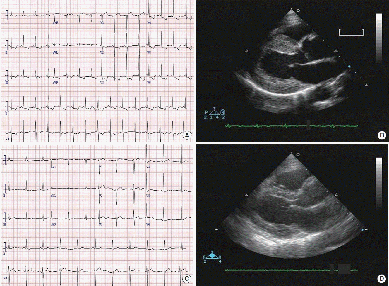
Electrocardiographic and echocardiographic findings. (A) Electrocardiography on admission day shows ST depression with T wave inversion in V2–6, lead II, III, and aVF. (B) Echocardiography on admission day shows left ventricular hypertrophy. (C) Abnormal electrocardiographic findings on admission day was normalized after surgery. (D) On follow-up echocardiography, left ventricular hypertrophy is disappeared after surgery.
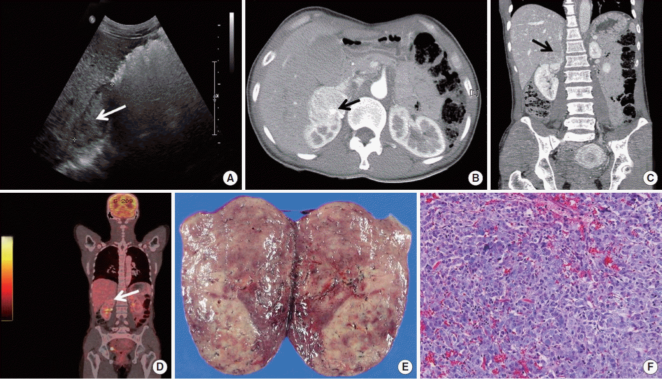
Imaging and biopsy findings. (A) Abdominal sonography shows about 6.15-cm hypoechoic mass-like lesion above the right kidney (white arrow). (B) Axial view on arterial phase (black arrow). (C) Coronal view on secretory phase. Abdominal computed tomography shows about 7-cm mass in right adrenal gland enhancing during arterial phase and washing out during secretory phase (black arrow). (D) 18F-fluorodeoxyglucose positron emission tomography shows about 5.0×3.2×6.5-cm-sized enhanced mass in right adrenal gland (maximum standardized uptake value 3.7) without metastasis (white arrow). (E) Gross specimen of pheochromocytoma in right adrenal gland shows about 7.0×4.0×2.0-cm-sized ovoid mass. The tumor is well-circumscribed and encapsulated. It is soft, yellow-tan to slightly brown. Focal hemorrhage and necrosis are shown. (F) On microscopic finding (H&E, ×200), tumor is composed of compact tumor cell nests surrounded by sustentacular cells and separated by fibrovascular stroma (zellballen appearance).
DISCUSSION
Pheochromocytoma is an uncommon neuroendocrine tumor originating from chromaffin tissue in adrenal gland and secreting catecholamines. The triad of typical symptoms is consisted of headache, palpitation, and diaphoresis [1]. Because catecholamines secreted by pheochromocytoma produces variant effect on the cardiovascular system, several alternative clinical manifestations other than classical triad of symptoms can lead to make a misdiagnosis [2].
Cardiovascular manifestations of pheochromocytoma include hypertension with or without left ventricular hypertrophy, myocarditis, cardiomyopathy, arrhythmia, and cardiogenic shock with pulmonary edema [3]. Of these, hypertension is the most common and important cardiovascular manifestation [2]. In addition, pheochromocytoma presents rarely as myocardial infarction. The mechanism of myocardial infarction in pheochromocytoma is due to a myocardial oxygen demand-supply mismatch. Coronary vasospasm, increased afterload with vasoconstriction, tachycardia, and a direct toxic effect by catecholamines can aggravate this mismatch resulting in chest pain, electrocardiographic changes (ST-T segment change), and increased cardiac enzyme in patients with normal coronary angiography [4,5].
Kim et al. [6] reported two cases of pheochromocytoma associated with acute myocardial infarction. In this report, increased cardiac enzyme, eletrocardiographic changes, and regional wall motion abnormalities on echocardiography were matched with acute myocardial infarction. But the results of coronary angiography were normal. Through the blood pressure fluctuation and paroxysmal increase in heart rate during admission pheochromocytoma was suspected and confirmed by adequate diagnostic process.
In our case, the patient’s clinical manifestation were compatible with myocardial infarction. Moreover, coronary angiography of the patient was normal. With careful history taking, patient’s symptoms attributed to perimenopausal syndrome which were eventually associated with underlying pheochromocytoma and blood pressure fluctuation were suspected for relating to pheochromocytoma and finally found to be compatible with it. This diversity of clinical manifestation of pheochromocytoma can delay the diagnosis and result deleterious cardiovascular outcome.
The treatment of choice in pheochromocytoma is surgical resection. Before surgery, α-blocking agent such as phenoxybenzamine should be preceded for at least 14 days. But in patients with pheochromocytoma presenting as acute myocardial infarction, β-blocking agents can be used because of myocardial infarction and result in catastrophic effects on pheochromocytoma due to an unopposed α-receptor stimulation aggravating patient’s outcome [5]. So our case emphasize the importance of considering pheochromocytoma as the differential diagnosis of patients with acute myocardial infarction without the evidence of coronary atherosclerosis on coronary angiography.
Pheochromocytoma is often fatal when doctors do not have a clinical suspicion as it can present as many other cardiovascular manifestations including acute myocardial infarction just like our case. In patients with acute myocardial infarction whose clinical feature cannot be exactly explained by the result of eletrocardiography, echocardiography, cardiac enzymes, and coronary angiography, doctors should have a high clinical suspicion for underlying pheochromocytoma, especially if there is blood pressure fluctuation. Early diagnosis and surgical treatment of an underlying pheochromocytoma can prevent the complication of this lethal but curable disease and result in good outcome without critical morbidity or mortality.