Rotational Atherectomy through Inner Guiding Catheter System for 1.25 mm Rotational Burr Non-Crossable Heavily Calcified Coronary Stenosis
Article information
Abstract
Among the various kinds of percutaneous coronary intervention techniques for balloon non-crossable severe calcified coronary stenosis, rotational atherectomy (RA) is known to be a therapy of choice. We describe a case in which a 1.25 mm RA burr non-crossable heavily calcified stenosis was successfully treated by the RA through ‘6 in 8 child-mother’ guiding technique.
INTRODUCTION
The development of various techniques and equipments for percutaneous coronary artery intervention (PCI) has led to an increase in age of the PCI population, and an increase in complexity of cases such as chronic total occlusion and calcified lesion. However, on the other hand, these subsets can increase the rate of balloon failure (inability to cross or dilate a coronary stenosis with a balloon) [1]. Among the many devices for PCI, rotational atherectomy (RA) has shown high procedural success rates with non-crossable calcified stenosis [2]. It is well known that RA for this frustrating subset is limited by the required size of guiding catheters and the ability to cross the lesion with a dedicated RotaWire [3], and 1.25 mm burr passage failure after successful RotaWire delivery is uncommon situation. In this report, we present a case in which 1.25 mm rotational burr non-crossable heavily calcified lesion was successfully treated by the RA through 6 in 8Fr inner guiding system.
CASE REPORT
An 85-year-old female patient was admitted to the coronary care unit due to resting chest pain and dyspnea with mild elevation of Troponin. Her coronary risk factors were hypertension and diabetes mellitus. The resting electrocardiogram showed ST depression in precordial leads, and echocardiography showed mild concentric left ventricular hypertrophy with a normal ejection fraction.
The emergency coronary angiography via the right femoral artery revealed diffuse moderate stenosis of the left anterior descending artery, and tight stenosis of the proximal left circumflex artery (LCX) and the distal right corona artery (RCA) with severe calcification (Fig. 1A-C). Firstly, we opened the proximal LCX lesion with a 3.0×14 mm Cilotax stent (Cardiotec Co., Seoul, Korea) without difficulty (Fig. 1D), and then we attempted to open the distal RCA lesion. After engagement of a 6Fr SH-JR 4 guiding catheter (Cordis Co., Miami, FL, USA) via the femoral artery, extra support choice PT guidewire (Boston Scientific Co., Marlborough, MA, USA) easily passed the lesion. But the smallest balloon available in our institution (1.2×6 mm Trek balloon; Abbott Vascular, Abbott Park, IL, USA) could not cross the lesion. The buddy wire technique was unsuccessful because any second (buddy) wires could not cross the lesion in the presence of the first wire. For stronger backup support, the guiding catheter was exchanged for a 6Fr SH-AL1.0 (Cordis Co.). But it also failed. Child-in-mother catheter technique (CMT) with a 5Fr 120 cm Heartrail ST01 (Terumo Co., Tokyo, Japan) was also unsuccessful for passing the balloon through the lesion. At this point, we realized that the RCA lesion was chronic severe calcified stenosis requiring a RA, and we stopped the procedure for second staged PCI with RA.
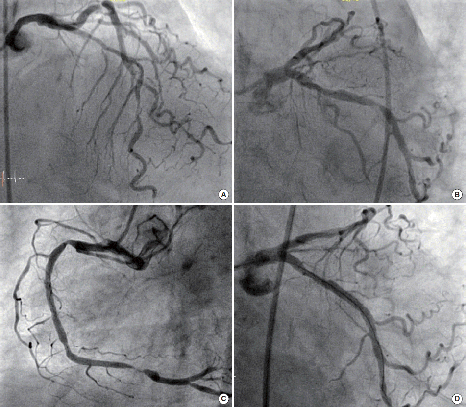
(A-C) Baseline images show diffuse moderate stenosis of the left anterior descending artery, and severe stenosis of the proximal left circumflex artery (LCX) and the distal right coronary artery with severe calcification. (D) The image after placement of a Cilotax stent at the proximal LCX.
After 7 days, we attempted second PCI with a 7Fr SH-AL1 guiding catheter (Cordis Co.) via the right femoral artery. Because we already observed ischemia during the CMT in the first PCI, a 3.5×18 mm Cilotax stent was placed at the proximal RCA stenosis, and then we tried again to cross the lesion using a Corsair microcather (Asahi Intec Co., Nagoya, Japan) and CMT with a 5Fr 120 cm Heartrail ST01, but without success (Fig. 2). As expected, the last resort was RA. Because the lesion did not allow two guidewire simultaneously, the first guidewire was removed and extrasupport Rota wire (Boston Scientific Co.) was then directly passed through the Heartrail ST01. After the Heartrail ST01 was removed, RA was tried to cross the lesion using a 1.25 mm burr (Boston Scientific Co.). But unfortunately, the burr could not cross the lesion despite any efforts. It seemed that there were no any further methods to overcome this situation. However, because of a possibility that RA pushing power was not sufficiently delivered to the distal target lesion, we decided to advance a Rotablator more distally toward the target lesion by using the CMT. A 100 cm 6Fr Envoy catheter (Cordis Co.) was used as an inner (child) catheter, and a 100 cm 8Fr SHJR4 catheter (Cordis Co.) was used as an outer (mother) catheter by shortening it to 80 cm (Fig. 3). By using the distal anchor balloon (2.5×20 mm) technique, the Envoy catheter could be advanced distally to make a RA-platform just proximal to the target lesion (Fig. 4A, B), which finally enabled the 1.25 mm burr to cross the lesion (Fig. 4C). After ablation was performed for 4 runs with a speed of 180,000 rpm, predilatations were easily performed using 2.0×10 mm and 2.5×20 mm balloons (Fig. 4D). Because following angiography showed long dissection, three Cilotax stents (2.5×28 mm, 3.0×28 mm, and 3.5×23 mm) were continuously implanted from the distal to the proximal RCA, yielding a good final result (Fig. 4E). After completing the procedure, the patient was stable without elevation of enzymes, and remained stable during the 8 months of clinical follow-up.

After a Cilotax stent was deployed at the proximal right coronary artery (RCA), (A) the attempt to cross the distal RCA lesion by the Corsair microcatheter and (B) deep intubation of a 5Fr 120 cm Heartrail ST01 was unsuccessful.

Images of 6-8 child-mother catheter system. (A) A 100 cm 6Fr Envoy catheter was used as an inner catheter, and a 100 cm 8Fr SH-JR 4 catheter was used as an outer catheter by shortening it to 80 cm with downsizing 7Fr sheath. (B) The distal tip of the Envoy catheter can protrude from the JR 4 catheter.

(A, B) By the distal anchor balloon technique, the inner guiding catheter (Envoy catheter) could be advanced just proximal to the target lesion. (C) 1.25 mm Rotablation was successfully performed through the inner guiding catheter. (D) Multiple predilataions were done through the inner guiding catheter. (E) Final angiogram after placement of three Cilotax stents.
DISCUSSION
Since the introduction of drug eluting stent (DES), the PCI for calcified lesions has increased because DES has shown a significant reduction in restenosis of both calcified and non-calcified lesions [4]. However, PCI for severely calcified lesions poses technical challenge. First, incomplete stent expansion can lead to the stent thrombosis or restenosis. Second, heavily calcified lesion sometimes does not permit stent or balloon passage which can lead to procedure failure. In order to overcome catheter passage failure in heavily calcified lesion, various techniques can be used: deep seating of guiding catheter, buddy wire technique [5], anchor balloon technique [6], use of microcatheter such as Tornusand Corsair (Asahi Intec Co.) [7,8], use of inner catheter (child mother technique) [9], and RA. Among these, RA is considered best option for this subset especially when other techniques are unsuccessful) [1,2].
In the present case, various techniques were applied for crossing this heavily calcified lesion, but without success even with 1.25 mm RA. The buddy wire technique (the second wire could not cross the lesion) and Tonus catheter (not available in our lab) could not be applied. We can also consider laser angioplasty with 0.9 mm X-80 catheter (not available in our country) [10]. However, considering the failure of 1.25 mm burr passage, these unused methods would be also unsuccessful. According to the previous RA studies for balloon non-crossable lesion, the most common cause of RA failure was inability to pass the dedicated RotaWire beyond the lesion [10,11], while the failure of RA burr passage after successful RotaWire delivery, as seen in the present case, is very rare. There can be some solutions for burr passage failure; (1) increasing the burr speed, (2) change of guiding catheter to more supportive one, (3) change of RA floppy wire to RA extrasupport wire, and (4) advance of RA platform very close to the target site, and etc. In this case, we focused that the target lesion was distally located with some distance from RA platform, and therefore we tried to advance Rotablator just near the target site in order to provide sufficient pushing power. But RA platform failed to go further beyond mid RCA segment. It is well known that the CMT is very useful technique for increasing back up support and delivering devices near or beyond the target lesion through tough proximal segment [9,12], which led to the application of CMT to RA in this case. The 1.25 mm burr passage through a 5Fr 120 cm Heartrail ST01 catheter (inner diameter is 1.5 mm) may be possible and there are several reports about RA using 5Fr guiding catheter [3]. However, considering both severe resistance during the delivery of the burr through a 5Fr catheter and previous 1.25 mm burr failure, RA through 5Fr inner guiding system would have a higher chance of failure than 6Fr inner guiding system for such a hard case. Therefore, for the purpose of smooth delivery of burr and stronger back up support, 100 cm 6Fr straight Envoy catheter originally designed for brain intervention was used as an inner catheter, and a 100 cm 8Fr JR-4 catheter was used as an outer catheter by shortening it to 80 cm because 6Fr catheter cannot enter the 7Fr catheter. Shortening of guiding catheter with a downsizing sheath (e.g., an 8Fr catheter with a 7Fr sheath) is simple maneuver which takes only 30 seconds, and sometimes used for retrograde approach for coronary chronic total occlusion with a very long collateral connection [13]. Also, this 6 in 8 CMT can be used in other frustrating situation such as vessel perforation where tough proximal anatomy frequently inhibits successful delivery of a graft stent with high profile [14]. To the best of our knowledge, this may be the first reported case, in which rotablation through the inner guiding system (6-8 CMT) was successfully done for 1.25 mm RA burr non-crossable heavily calcified stenosis. However, disadvantages of this technique should not be disregarded. Because a relatively large bore inner catheter have to pass through a proximal segment, it is generally impossible to reach the target site without anchor balloon technique especially when the proximal segment has a disease. Therefore, operators should consider the possibility of a dissection in the proximal segment, which can lead to protracted length of stenting, and ischemia especially in the case of multivessel disease. Although there can be a doubt for how many cases the RA through the CMT can be applicable, it certainly helps to know this novel technique when we encounter a very tough calcified lesion.