Laparoscopic Removal of Gossypiboma Following Cesarean Section
Article information
Abstract
Gossypiboma refers to a tumorus mass that is caused by a foreign body reaction due to an accidentally retained gauze or sponge within the body following surgery. Here, we report the case of gossypiboma in a 29-year-old woman with lower abdominal discomfort over a period of 2 years. In her medical history, she had a Cesarean section 11 years ago. Transvaginal ultrasonography showed an 8.5×6.4 cm mixed echoic round mass and both adnexa were normal. Abdominal-pelvic computed tomography confirmed a high attenuation lesion with a suspected foreign body in the pelvic cavity. Thus, based on a diagnosis of gossypiboma, she received a laparoscopic operation. Upon laparoscopic view, the mass was discovered to be well encapsulated and solid, and located between the urinary bladder and uterus. The mass was completely removed without damage to other adjacent organs. The patient was discharged on the 5th postoperative day without complications.
INTRODUCTION
Gossypiboma refers to a tumorous mass that is caused by a foreign body reaction due to an accidentally retained gauze or sponge within the body following surgery. Despite of careful intraoperative precautions and gauze counts, mistakes that can cause significant morbidity still occur and may result in death [1]. Clinical symptoms of gossypiboma are highly variable, therefore, gossypiboma should be considered as a differential diagnosis in any postoperative patients with pain, infection, or a palpable or asymptomatic mass that is incidentally discovered in the same anatomic area. However, prevention is most important than an accurate diagnosis and adequate treatment. Here, we report the case of a gossypiboma in a 29-year-old woman who had a Cesarean section 11 years ago.
CASE REPORT
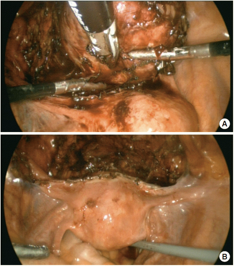
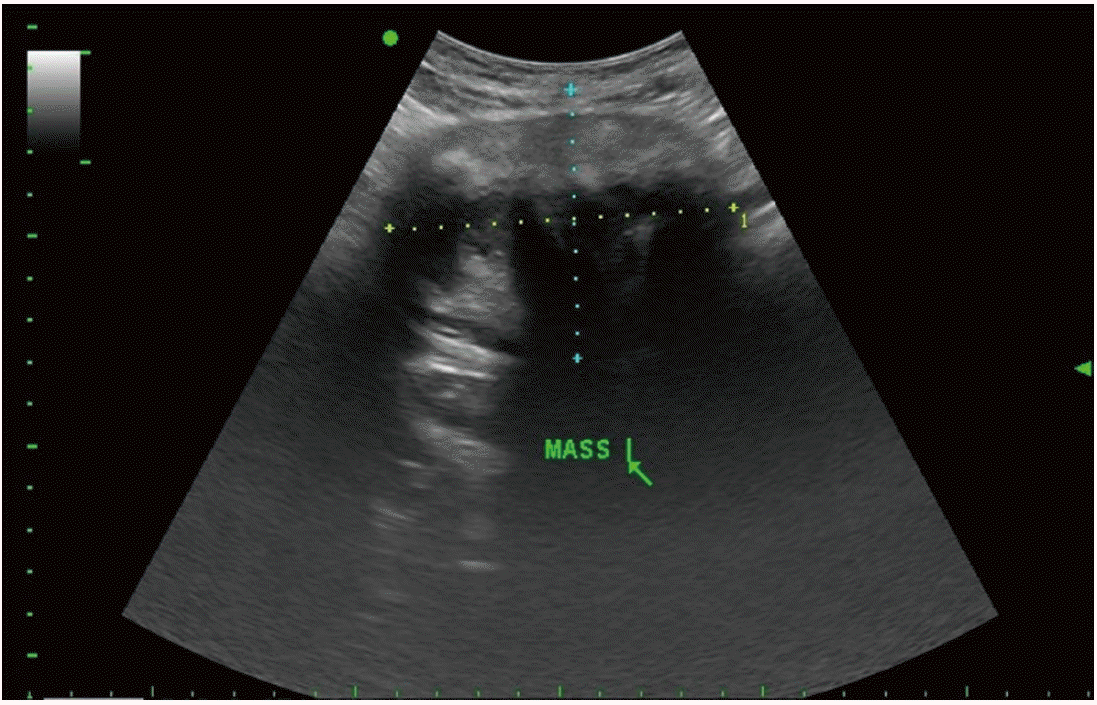
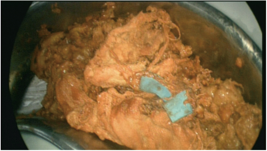
A 29-year-old Asian woman visited Soonchunhyang University Cheonan Hospital with lower abdominal discomfort over a period of 2 years. She had been diagnosed with pelvic inflammatory disease and treated but symptoms had not improved. During follow-up, a pelvic tumorous mass was confirmed on transvaginal ultrosonography (USG) at a local clinic. The patient was referred to Soonchunhyang University Cheonan Hospital for further evaluation and treatment. According to her medical history, she had a Cesarean section 11 years ago. Her initial vital signs were as follows: blood pressure, 120/80 mm Hg; pulse rate, 76 beats/min; respiratory rate, 20 beats/min; and body temperature, 36.5°C, without unusual findings. Peripheral blood tests showed a hemoglobin level of 14.7 g/dL, white blood cell level of 5,030/mm3, and platelet count of 340,000/mm3. Biochemistry and urine analyses were all within the normal range. Additionally, titers of the tumor markers cancer antigen 125 and carbohydrate antigen 19-9 were within the normal range. Transvaginal USG showed an 8.5×6.4 cm mixed echoic round mass (Fig. 1) and both adnexa were normal. Plain abdominal radiographs were interpreted by a radiologist as normal (Fig. 2). Abdominal-pelvic computed tomography (CT) confirmed a high attenuation lesion with a suspected foreign body in the pelvic cavity (Fig. 3). Thus, based on a diagnosis of gossypiboma, she received a laparoscopic operation. Upon laparoscopic view, the mass was discovered to be well encapsulated and solid, and located between the urinary bladder and uterus (Fig. 4A). The mass was completely removed without damage to other adjacent organs (Fig. 4B). Upon gross examination (Fig. 5), the specimen was sliced into many fragments and a surgical sponge was visible to the naked eye. On pathologic examination, the final diagnosis was foreign body granuloma. The patient was discharged on the 5th postoperative day without complications.

Abdominal-pelvic computed tomography scan. Mass with internal radiopaque material (arrow) was seen in pelvic cavity.

Intraoperative laparoscopic view. (A) Encapsulating solid mass located in front of uterus. (B) Laparoscopic finding after removal of mass.
DISCUSSION
Every operation with an incisional wound carries the potential risk of retained foreign bodies, such as a surgical sponge, needle or instrument. Real instrument counts are a very important for safety during operations. However, these procedures do not exclude the possibility of a retained surgical sponge. Approximately 77 to 88% of cases with retained foreign bodies occur in operations with correct counts [2-4]. If an incorrect count occurs, a portable radiograph is occasionally used to identify foreign bodies within the human body, and this event is not rare in the operating room in Soonchunhyang University Cheonan Hospital. Among foreign bodies, surgical sponge is most frequently retained in the human body after an operation and can cause gossypiboma [3]. Gossypiboma is derived from the combination of the Latin word ‘gossypium’ meaning cotton and the Swahilli word ‘boma’ meaning place of concealment and refers to a lesion resulting from a retained gauze or sponge after an operation [5]. Although the incidence of gossypiboma is unknown in South Korea, the incidence varied from 1/8,801 to 1/18,760 inpatient operations in the United States [3]. However, previous studies suggest that the incidence of gossypiboma has been underestimated and rarely discussed because of legal implications. Another study reported the real incidence of retained foreign bodies to be 1 in 5,500 inpatient operations and the author suggested that this complications may occur more frequently than expected from literature [4].
Among the body cavities the abdomen or pelvic cavity was most commonly involved by remaining foreign bodies [2,3,5]. Many authors have suggested that risk factors for retained foreign bodies are associated with multiple major surgeries, emergency surgeries, unplanned changes in the operation and, in obese patients, longer operations with increased complexity [2,3]. In high risk patient with gossypiboma, we should approach with great care including radiograph scanning before leaving the operating room even when sponge counts have been deemed to be correct.
Clinical symptoms of retained foreign bodies are highly variable depending on the location and types of foreign body reactions. The first type of reaction is exudative in nature and usually occurs shortly after surgery. In these cases, symptoms are caused by abscess formations. In addition, intestinal obstruction and fistula formation may occur. The second type is an aseptic fibrous response that may create adhesions and encapsulation via a foreign body granuloma that, as in our case, would not be detected for a long time [6].
Every gauze and sponge used during an operation in South Korea now contains radiopaque markers for detection by radiography. Therefore, conventional plain radiographs theoretically should be able to detect gossypiboma. However, these markers can be easily overlooked, as they become distorted by folding or twisting, and may even disintegrate over a period of time [2]. Additionally, if a radiologist has no clinical information regarding the patient and has no knowledge and experience concerning gossypiboma, this mass can be easily overlooked. In our experience, although radiopaque markers have not been integrated in plain abdominal radiograph and the curved radiopaque line can be easily observed, a radiologist can overlook the foreign body, resulting in a false-negative finding. A typical USG finding of gossypiboma demonstrates a well-defined hypoechoic mass containing highly echogenic foci with a strong posterior shadow [7]. However, our case did not show that typical form of gossypiboma, but showed a mixed echoic lesion that was misdiagnosed as a neoplasm instead. Abdominal CT is the most effective and practical diagnostic method for gossypiboma [5]. Although the CT findings of gossypiboma are various, the most specific finding is a soft tissue mass with air bubbles and a spongiform pattern [2]. In our case, abdominal CT did not show a typical gossypiboma pattern, but instead demonstrated a round, well-encapsulated mass. Internal radiopaque material was noted and it was therefore preoperatively diagnosed as a foreign body granuloma. Curative treatment of gossypiboma implies removal of the gossypiboma either by open or laparoscopic approach [1]. The current recommendations suggest that laparoscopic approach could be attempted in recent, small inflammatory pseudotumors, with obvious encapsulation and no complications [8]. However, in this case, a gossypiboma was completely removed using laparoscopic surgery without damage to other adjacent organs, although it was a longlasting retained large-sized mass.
In conclusion, although a diagnosis of gossypiboma is difficult because the clinical symptoms are highly variable, gossypiboma should be considered as a differential diagnosis for any postoperative patient who presents with pain, infection, or a palpable mass or an asymptomatic mass that is incidentally discovered in the same anatomic area. However, the best treatment for this complications is prevention rather than accurate diagnosis and adequate treatment. In high risk patient with gossypiboma, we should approach with great care.


