Acute Myocardial Infarction with Simultaneous Occlusions of Left Anterior Descending Artery and Right Coronary Artery
Article information
Abstract
Acute myocardial infarctions involving multiple coronary arteries simultaneously are infrequent and causative risk factors of the occlusions are unclear. However, severe complications arise, such as congestive heart failure or death. We report a case of two simultaneously occluded coronary arteries. A 39-year-old Korean man with simultaneous total occlusion of the left anterior descending artery and the right coronary artery presented with chest discomfort and cardiogenic shock. Immediate percutaneous coronary intervention was performed and a transvenous temporary pacemaker and intra-aortic balloon counterpulsation catheter were inserted. Through continuous effort he was discharged 8 days post intervention without any complaints.
INTRODUCTION
Acute myocardial infarction (AMI) involving two or more coronary arteries simultaneously has worse prognosis than a single artery occlusion regarding a high risk of death [1-3]. We report a 39-year-old Korean man with simultaneous total occlusion of the left anterior descending artery and right coronary artery, presenting with chest discomfort, cardiogenic shock, ventricular tachycardia and finally asystole. After successful percutaneous coronary intervention (PCI), the patient was discharged 8 days post intervention.
CASE REPORT
In November 2012, a 39-year-old Korean male was referred to the emergency room (ER) after losing consciousness. While he was playing baseball, he felt chest pain and soon fainted. When his friends noticed that he was pulseless, they started cardiac massage for about 5 minutes, which led to the regaining of his pulse. A physical examination showed that his respiratory rate was 20/min, his heart rate was 114/min, and his blood pressure was 80/60 mmHg. An electrocardiogram (ECG) was immediately applied and showed ST elevation in the inferior (II, III, and aVF) leads and anterior (V1-V6) leads, and a Q wave in the anterior (V1-V3) leads (Fig. 1), which was suggested inferior and anterior AMI. Smoking was the only risk factor for coronary artery disease. A chest X-ray revealed no pulmonary congestion. The patient was given 300 mg of aspirin, 600 mg of clopidogrel and an intravenous heparin infusion as per primary PCI protocol, while waiting for emergent PCI. Initial laboratory data showed Troponin-I <0.10 ng/mL (range, 0.00 to 0.16 ng/mL), creatinine kinase (CK) 193 IU/L (range, 56 to 244 IU/L), CK-MB isoenzyme 3.2 ng/mL (range, 0.0 to 5.0 ng/mL), myoglobin 164 ng/mL (range, 0 to 72 ng/mL), and glucose 437 mg/dL (range, 70 to 100 mg/dL). When the patient arrived at the angiography room, his heart rate was 40 beats/min and a temporary transvenous pacemaker was inserted (VVI 70 bpm, 10 mA, and 2.0 mV).
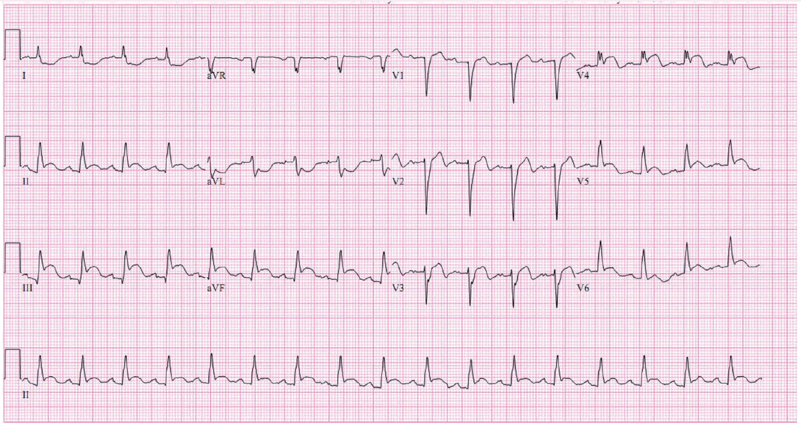
Initial electrocardiogram on arrival shows ST elevation in the inferior (II, III, and aVF) leads and anterior (V1-V6) leads, and a Q wave in the anterior (V1-V3) leads.
Coronary angiography revealed two-vessel coronary disease, with a thrombus and total occlusion of the middle right coronary artery (RCA) (Fig. 2A) and proximal left anterior descending artery (LAD). A minimal stenosis at the left main coronary artery was also observed (Fig. 2B). There was no significant luminal narrowing of the left circumflex artery. We could not find any collateral circulation to the RCA or LAD. An intervention was performed first on the middle RCA. The lesion was crossed with a Runthrough NS (Terumo, Tokyo, Japan) guidewire easily and smoothly. After the balloon was dilated, a thrombus formation was revealed. An angioplasty was performed with an implantation of a 2.75×18 mm Resolute integrity (Medtronic, Galway, Ireland) stent (Fig. 2C). There was neither slow-flow nor no-reflow in the RCA after the intervention. However, there was no collateral circulation from the RCA to the LAD and an intervention at the proximal LAD lesion was also performed. The guidewire for LAD lesion was also easily and smoothly passed. An implantation with a 2.75×22 mm Resolute integrity stent followed by balloon dilatation was done at the proximal LAD (Fig. 2D). We did not administer intravenous glycoprotein (GP) IIb/IIIa inhibitor as the thrombus was not seen in the culprit lesions after the final intervention.
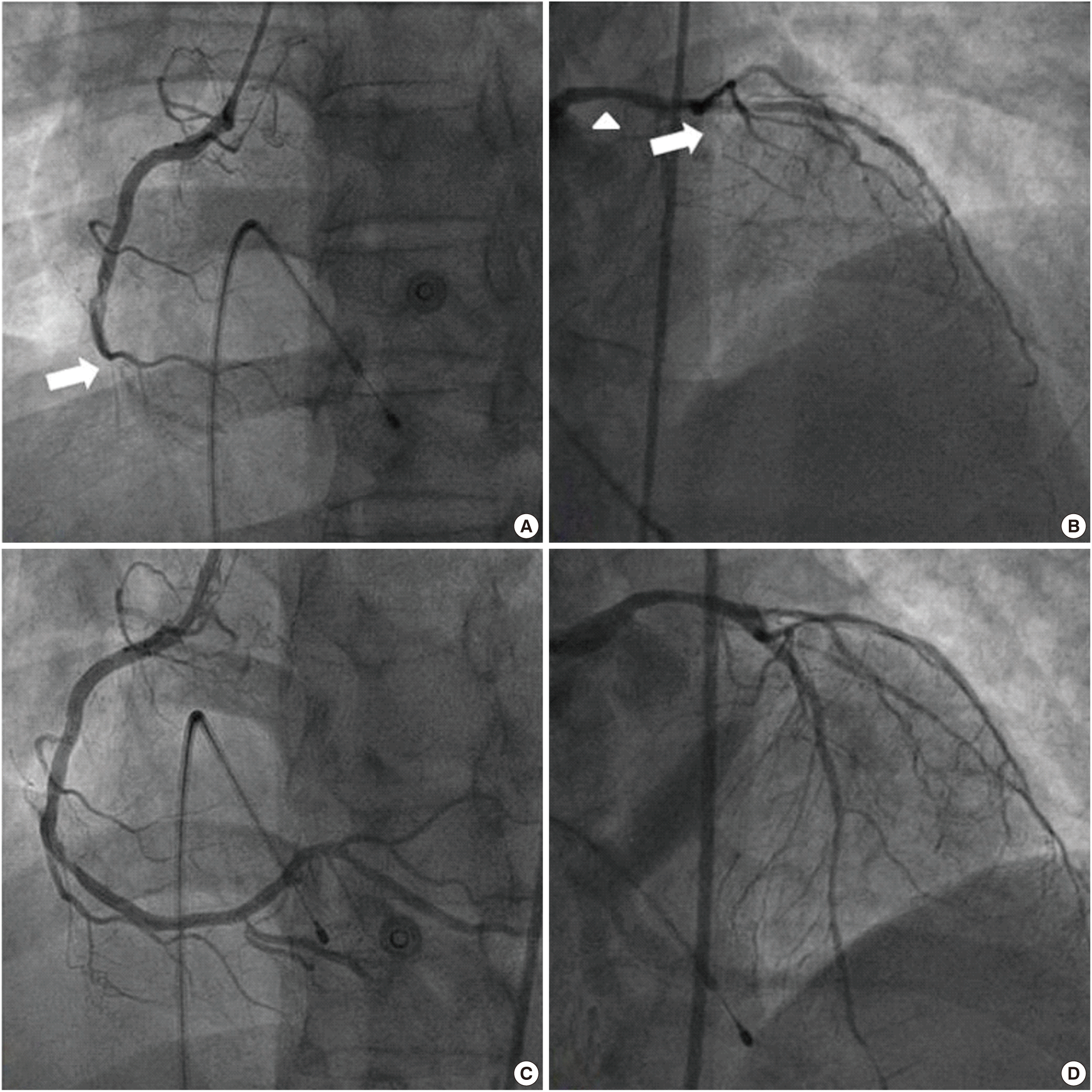
Emergent coronary angiograms during percutaneous coronary intervention. (A) Total occlusion and thrombus formation (arrow) at the middle right coronary artery (RCA). (B) Total occlusion and thrombus formation (arrow) at proximal left anterior descending artery (LAD) and minimal stenosis (arrow head) at the left main coronary artery. (C, D) After intervention, no residual stenosis at the RCA and at the LAD.
The blood pressure was 60/40 mmHg after the PCI, when an intra-aortic balloon counterpulsation (IABP) was introduced (1:1 triggered by ECG). However, about 10 minutes after the PCI, all of a sudden the patient lost consciousness with ECG alterations, notably in the ventricular tachycardia and then entered asystole. Cardiopulmonary resuscitation with cardioversion was initiated and a spontaneous rhythm of the patient was recovered. After removal of IABP and the pacemaker, 2 days after admission, the patient had no sign of recurrence of chest pain and was electrically and hemodynamically stable in New York Heart Association class II, and recovered to the normal state of consciousness, maintaining good urine output, without evidence of ventricular arrhythmias on the monitor. To find out other co-existing diseases which could create a thrombus formation, we examined the chest computed tomography (CT) and blood tests including anti-phospholipid immunoglobulin G (IgG), anti-cardiolipin IgG, anti-cardiolipin immunoglobulin M, lupus anticoagulant, anti-β2 glycoprotein IgG, protein C activity, protein S activity, homocysteine and anti-thrombin III. The examination results showed no thrombus on the chest CT or any abnormality in the blood tests. The echocardiogram after the procedure showed moderate dysfunction of the left ventricular systolic function with a mean ejection fraction of 37%, akinesia of the mid to apical anteroseptal wall, left ventricle apex and basal inferior wall, and severe hypokinesia of the mid inferior wall. Eight days later, he was discharged home without chest pain or pulmonary congestion. The ECG at the time of discharge showed no ST elevation (Fig. 3). The patient is free of symptoms and is visiting the out-patient clinic prescribed with aspirin, clopidogrel, statin, angiotensin-receptor blocker, beta-blocker, furosemide, and spironolactone.
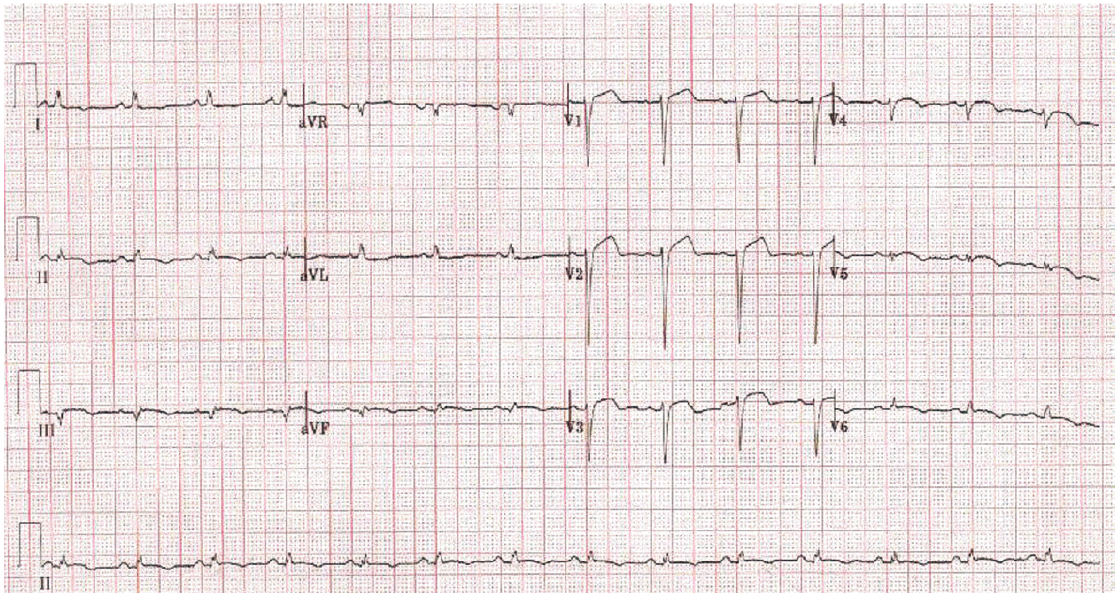
Electrocardiogram before discharge shows a remaining Q wave in the anterior leads (V1, V2 and V3), but no ST elevation in the inferior leads (II, III, and aVF) and anterior leads (V1-V6).
DISCUSSION
The main causes of the ST segment elevation myocardial infarction were the plaque rupture and the subsequent thrombosis of a single culprit vessel. However, simultaneous occlusions involving more than 2 coronary arteries are not due to local plaque pathology. The causative factors involved in the acute and simultaneous thrombosis of multiple coronary vessels are unclear; there are some reported possible causative factors. These are hemodynamic instability and hypotension due to occlusion of one coronary artery, resulting in blood stasis and acute occlusion in another artery with a severe underlying lesion [4,5], hypercoagulable states, such as malignancy or thrombocytosis [6,7], and antithrombin III deficiency [8]. However, when a patient comes to the ER with the impression of AMI, it is very difficult to verify these causative factors. Therefore early administration of an ECG and timely angiography are important. In this case, an initial ECG taken in the ER showed ST elevation of two coronary vessel territories (inferior and anterior) which made us suspect double vessel occlusion and the following angiography confirmed this speculation. As the patient was already in cardiogenic shock, thrombolysis was not indicative. An emergency PCI for the RCA was primarily done since the patient displayed bradycardia. After successful intervention of the RCA, no-reflow or slow-flow was not observed and collateral circulation to the LAD was not found, therefore we decided to perform a PCI at the LAD lesion, also. As both lesions were passed by guidewire easily and smoothly and there was no collateral circulation from other vessels to the RCA or the LAD, we are sure it is the case of simultaneous double vessel occlusion. After coronary intervention of both the LAD and RCA lesions, neither no-reflow nor slow-flow was observed and residual thrombus was not seen and therefore we did not use GP IIb/IIIa inhibitor. However, we feel at odds about the occurrence of cardiac arrest which might have been prevented by using GP IIb/IIIa inhibitor. This patient had only one risk factor—smoking. Further, there were no laboratory abnormalities which could cause coagulation problems or other systemic conditions such as a pulmonary embolism. In this case, a hemodynamically unstable condition such as cardiac arrest, before arriving at the hospital might have attributed to his simultaneous double coronary artery occlusion.
An AMI with simultaneous two or more vessels occurs rarely but has a poorer prognosis than a single vessel disease [6,8]. In one review article, more than 50% of the patients in the previous reports suffered from cardiogenic shock [9]. In addition, an autopsy series in patients who died from AMI report that a thrombotic occlusion of more than one major coronary artery is not rare, occurring in up to 50% of the patients [10]. This may suggest the fact that AMI with multivessel occlusion often leads to massive myocardial damage and death before the patient arrives at the hospital. Fortunately in this case, the patient came to hospital immediately and intervention was done promptly. Also, treatment using an IABP was effective enough to allow the patient to be discharged soon after gaining back normal vital signs. Therefore, an early diagnosis and proper management are the most important things in the therapeutic course.
In conclusion, patients with multiple occlusive coronary artery disease are often in a devastating condition. Cautious attention must be given for the accurate diagnosis in an abnormal ECG showing ST segment elevation. Further, early diagnosis and proper management including timely intervention are the most important things in the therapeutic course.