Endometriosis of the Appendix
Article information
Abstract
Endometriosis is a well-recognized gynecological condition in the reproductive age group. Endometriosis of the appendix is an entity of extragonadal endometriosis. The incidence of appendiceal endometriosis is lower than 1% among pathologies of pelvic endometriosis. Women can present with symptoms mimicking acute appendicitis or chronic pelvic pain. Diagnosis can be made only after a histopathological examination following the operation. We report here a case of appendiceal endometriosis, which were operated on for a prediagnosis of acute appendicitis, but postoperatively diagnosed as appendiceal endometriosis.
INTRODUCTION
Endometriosis is defined as the presence of ectopic endometrial tissue outside the lining of the uterine cavity [1]. Endometriosis is a common condition that can affect up to 15% of women of child-bearing age and 2% to 5% of post-menopausal women [2]. Endometriosis is seen at a rate of 3% to 37% in various parts of the gastrointestinal system, from the small intestine to the anal canal and 76% of these occur in the sigmoid colon and rectum [3]. Appendiceal endometriosis is a rarely seen condition comprising less than 1% of pelvic endometriosis cases [4] and preoperative diagnosis is difficult. In the present study, we report a case of appendiceal endometriosis, which we operated on for a diagnosis of acute appendicitis.
CASE REPORT
A 45-year-old woman was admitted with a 1-day history of right lower-quadrant pain and nausea. Her past medical history was significant for twice cesarean section and subtotal hysterectomy for uterine myoma. The physical examination revealed tenderness and positive rebound in the right lower abdominal quadrant. No abdominal mass was palpable. Axillary body temperature was 36.4°C. The patient’s white blood cell count was 12,950/mm3 with 84.1% neutrophils and C-reactive protein was 1.89 mg/dL. Urine analysis was normal, with no evidence of infection or haematuria. She was evaluated with an abdominal and pelvic computed tomography (CT), which demonstrated a mild dilated appendix with enhancing wall thickening and minimal low density fluid collection in periappendiceal space of right lower abdominal quadrant. Based on the clinical and laboratory findings, we suggest the diagnosis of acute appendicitis.
After acute appendicitis was diagnosed, a laparoscopic appendectomy was performed. Intraoperatively, the appendix appeared mildly congested and edematous. Minimal reactive fluid in Douglas pouch was determined. There is no abnormal finding in pelvic cavity.
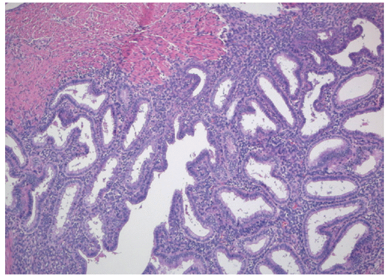
Postoperative course was uneventful and the patient was discharged the third postoperative day. The appendix measured 3×2.5 cm at the widest diameter. Grossly, the serosal surface shows increased vascularity. Histopathologic examination demonstrated acute appendicitis and endometrial glands with stroma in the thickened muscularis propria of the appendix, so the pathology report led to the diagnosis of appendiceal endometriosis (Figs. 1, 2). The postoperative gynecological examination did not reveal any other endometriotic lesions.
DISSCUSSION
Endometriosis is a condition characterized by the growth of the endometrial tissue outside the uterine cavity. Endometriosis is commonly found in the genital organs and the pelvic peritoneum. The most common sites of endometriosis involvement are the ovaries (54.9%), posterior broad ligament (35.2%), anterior cul-de-sac (34.6%), the posterior cul-de-sac (34.0%), and the uterosacral ligament (28.0%) [5]. Involvement of the gastrointestinal tract is reported to affect between 3% and 37% of patients with pelvic endometriosis [3]. Involvement of the gastrointestinal tract is uncommon. When endometriosis does involve the gastrointestinal tract it commonly involves the recto-sigmoid colon (72%), the recto-vaginal septum (13%), small intestine (7%), cecum (3.6%), and the appendix (3%) [6].
Appendiceal endometriosis patients can be categorized into four groups in terms of symptomatology: 1) patients who present with acute appendicitis; 2) patients who present with appendix invagination; 3) patients manifesting atypical symptoms such as abdominal colic, nausea and melena; and 4) patients who are asymptomatic [7]. The most commonly seen group comprises patients who present with appendicitis. Acute appendiceal inflammation can arise because of partial or complete luminal occlusion by the endometrioma [8]. Another mechanism suggested is that of endometrium hemorrhage within the seromuscular layer of appendix, which is followed by edema, obstruction and inflammation. Pain in the right lower abdominal quadrant is one of the most common symptoms of appendiceal endometriosis. Although laboratory results are not specific, leukocytosis along with subfebrile fever is mostly present. Leukocytosis with the predominance of polymorphonuclear leukocytes accompanies acute appendicitis in most cases, along with elevated C-reactive protein. In our patient, fever was absent, but there was an increase in leukocytes. CT scans obtained to diagnose appendiceal endometriosis often show a distended, nonopacified appendix without inflammation [9]. In our patient who present with acute appendicitis, CT scans reveals a mild dilated appendix with enhancing wall thickening.
Because endometriosis of the appendix can manifest in many ways without any specific indications, it is difficult to make an accurate preoperative diagnosis. Laboratory tests are of limited value. CT of the abdomen and pelvis may show evidence of acute appendicitis, or appendiceal abnormality. Laparoscopy is considered the gold standard for the diagnosis of endometriosis. Pain is the most common indication for surgical management. The correctly diagnosis of appendiceal endometriosis is only established by the histological presence of endometrial tissue in the specimen [8].
About half of endometriosis of the appendix involves the body and half involves the tip of the appendix. Glandular tissue, endometrial stroma and hemorrhage are typical examinations conducted in patients with endometriosis [6]. Muscular and seromuscular involvement occurs in two-thirds of patients, while the serosal surface is involved in only one-third of patients. The mucosa is not involved, but Langman et al. [10] found that the submucosa was involved in one-third of patients with endometriosis of the appendix. In our patient, endometrial glands and stroma are found in muscular propria of appendix, too.
The treatment consists mainly of surgery and hormone therapy. The treatment tends to be determined by the age of the patient and the degree of the patient’s symptoms. A gynecological assessment should be performed to determine the extent of endometriosis. At laparotomy or laparoscopy, a careful examination of the abdominal cavity is carried out in order to fully evaluate the extent of disease. Postoperative follow-up is mandatory for appendiceal endometriosis. Medical treatments for endometriosis are secondary. The postoperative gynecological examination did not reveal any other endometriotic lesion in our patient and she had normal serum CA125 (cancer antigen 125) level, so it is not necessary for her to receive additionally treatment.
Endometriosis of the appendix is a rare situation that can simulate an acute appendicitis, and its preoperative diagnosis is difficult. However, it should be included in the differential diagnosis of acute abdominal pain, especially when women of childbearing age present with clinical symptoms of acute appendicitis but no evidence is observed on imaging studies. Laparoscopy is useful for the diagnosis, and appendectomy relieves the acute symptoms. Surgeons also need to discuss whether to apply elective appendectomy on patients who had undergone gynecological operation because of endometriosis.

