크기가 증가하여 전이성 병변으로 의심된 간자색반병 1예
A Case of Peliosis Hepatis Mimicking Metastatic Lesions of Increasing Size
Article information
Trans Abstract
Peliosis hepatis, an uncommon vascular condition, is characterized by multiple blood-filled cavities distributed throughout the liver. Computed tomography and magnetic resonance imaging findings of peliosis hepatis are nonspecific. A 40-year-old woman presented with multiple hepatic cystic masses. Two years later, the number and sizes of the masses had increased. We suspected meta-static hepatic disease and performed a liver biopsy. Histological examination revealed dilatation of hepatic sinusoids and multiple blood-filled cavities throughout the liver parenchyma. Thus, a diagnosis of peliosis hepatis was confirmed.
서 론
간자색반병(peliosis hepatis)은 간실질 내에 혈액이 차있는 여러개의 낭성질환으로 정의한다. 간자색반병은 대개 증상이 없어 우연히 발견되는 경우가 대부분이다. 국내에도 보고된 바 있으며 대개의 경우 조영증강 전 컴퓨터단층촬영(computed tomography, CT)에서 다발성의 저음영 또는 낭성 병변으로 보이고 조영증강 후 CT에서 병변의 크기, 굴모양혈관과 연결 정도, 혈전이나 출혈 등에 따라 다양하게 조영증강된다. 원심성 조영증강양상은 정맥확장형 간자색반병에서만 볼 수 있는 특이소견이나[1], 대부분 간자색반병의 영상소견은 매우 다양한 것으로 알려져 있다. 그러나 추적관찰에서 종괴의 크기가 증가한 보고는 없었다. 본 증례에서는 조영증강지연기까지 저조영증강으로 관찰되고 일부 테두리 조영증강의 모습이 보이며 종괴의 크기가 이전보다 증가한 간의 다발성 종괴를 전이성 병변으로 의심하여 간조직 생검을 통해 병리적으로 확진된 간자색반병 1예를 보고하는 바이다.
증 례
순천향대학교 부천병원에서 4년 전 왼쪽 유방의 생검절제술로 경화샘증을 진단받고, 2년 후 건강검진에서 간의 다발성 종괴가 발견되어 영상학적 검사와 조직검사를 통하여 지방 변성(macro-vesicular fatty change)으로 진단받았던 41세의 여자가 다발성 종양의 크기가 커져 내원하였다. 내원 당시 약제복용의 기왕력은 없고 특이 호소증상은 없었으나 간역동 CT 검사를 시행한 결과 다발성 종양의 크기가 2년 전보다 증가되어 있었다.
당시 복부초음파 검사결과 간 전체에 걸쳐 일부 얇은 고에코 테두리에 둘러싸여 있는 무에코 내지는 저에코 종괴들이 보였고 대부분 1-2 cm 내외로 관찰되었다(Fig. 1). 간역동 CT를 시행하였을 때, 조영증강 이전 영상에서는 여러 개의 저음영 종괴가 간 전반에 걸쳐 퍼져있고 대부분 조영증강이 거의 되지 않았다(Fig. 2A-D).
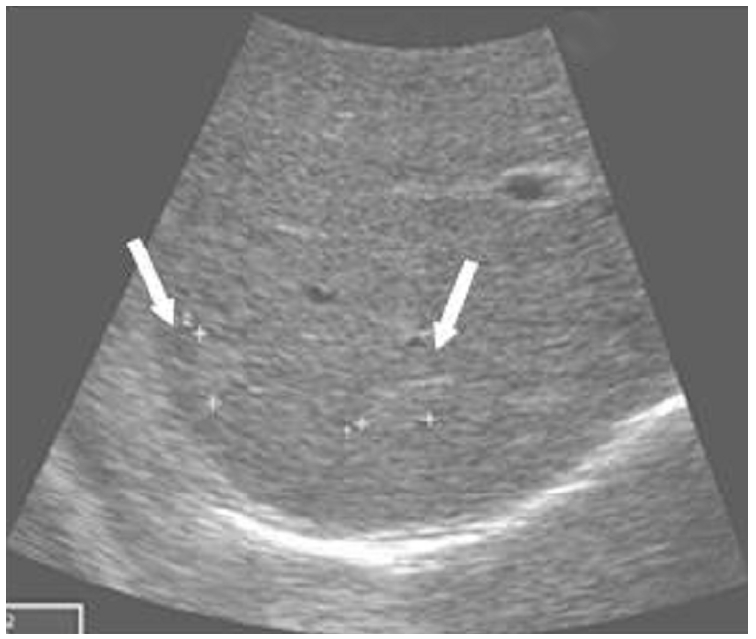
Ultrasonography. Hypoechoic mass is surrounded by a thin hyperechoic rim in the liver (2 years ago).
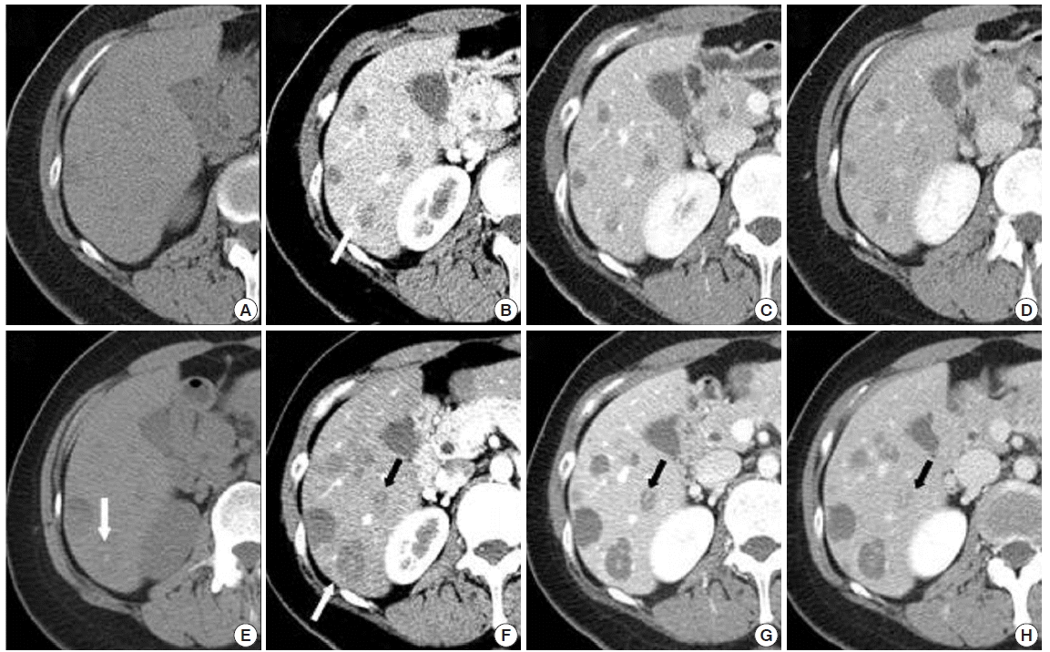
Liver dynamic computed tomography (CT). The precontrast phase (A, E), hepatic arterial phase (B, F), venous phase (C, G), and delayed phase (D, H). Masses displayed hypoattenuation in relation to liver parenchyma (A–D). The 2-year follow-up CT showed multiple mass size increases (E–H). The largest increase was from 8.9 to 21.6 mm (B, F, white arrow). Some masses were newly developed, and some contained calcification (E, white arrow). Masses were hypoattenuated on non-enhanced CT (E); in the arterial phase, lesions appear hypoattenuated in relation to the liver (F) and progressively became hyperattenuated (G, H).
생징후는 안정적이었고, 촉진상 유방의 종괴와 두경부와 액와 및 서혜부에 림프절도 없었고, 복부 오른편의 간비대 및 압통이나 종괴 또한 없었다. 혈액검사에서 혈청 aspartate aminotransferase(AST) 17 IU/L, alanine aminotransferase (ALT) 19 IU/L, 총 빌리루빈 0.4 mg/dL로 정상이었고, 종양표식자인 alpha fetoprotein, carbohydrate antigen 19-9 모두 정상이었다. 추적검사로 시행한 간역동 CT에서는 2년 전 시행한 CT와 비교하여 간에 있던 여러 개의 저음영 종괴들은 대부분 그 크기가 커졌다. 종괴의 크기는 가장 큰 차이가 직경 8.9 mm에서 21.6 mm로 크기가 증가하였으며 대부분 직경이 2배 가량 증가하였다. 경계는 비교적 좋은 편이고 일부는 약하게 조영증강되며 지연기에서 좀더 조영증강되었다(Fig. 2E-G). 일부는 이전 CT에서는 보이지 않았던 석회화들이 새로 생긴 상태였다(Fig. 2E). 자기공명영상(magnetic resonance imaging, MRI) T1강조영상에서는 경계가 불분명한 저신호강도로, T2강조영상에서는 주위 간실질보다 고신호강도로 보였다(Fig. 3A, B). 조영증강 지연영상에서는 일부는 주변 실질과 같은 신호강도를 보이고, 일부는 테두리 조영증강을 보이며 일부는 저조영증강으로 보였다(Fig. 3C). Positron emission tomography-CT에서는 비정상적인 fludeoxyglucose (FDG) 섭취 증가는 관찰되지 않았다(Fig. 4). 간의 다발성 종괴들의 크기가 2년 전에 시행한 검사보다 증가함에 따라 악성을 감별하기 위하여 초음파유도하 생검을 진행하였다. 조직검사결과, 굴모양혈관의 확장과 더불어 간실질 내에 여러 개의 혈액낭이 관찰되어 간자색반병에 합당한 소견이었으며(Fig. 5), 환자는 현재 간자색반병으로 진단받은 후에 별다른 치료없이 추적관찰 중이다.

Magnetic resonance imaging (MRI). (A) T1-weighted MRI shows an iso-intense and subtly hypointense lesion (arrow) compared with the surrounding liver parenchyma. (B) T2-weighted MRI shows a lesion of high signal intensity (arrow). (C) T1-enhanced MRI shows a low-enhancement lesion (arrow).
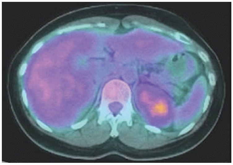
Positron emission tomography-computed tomography. No abnormal metabolic activity was detected in liver.

Pathologic findings (H&E, × 200). The liver tissue contains irregular, blood-filled, cystic dilatations of sinusoidal spaces.
고 찰
간자색반병은 간실질 내에 혈관 이상으로 생기는 다발성 낭성 종양으로 낭 내에는 혈액으로 차있다. 낭의 크기는 대개 mm에서 3 cm 정도이며 간실질 내 어디든 생길 수 있다. 대부분 증상이 없으나 황달, 간비대, 간부전 및 심한 경우 파열로 인한 복강 내 출혈이 나타나기도 한다.
간자색반병은 스테로이드, 안드로겐, 경구피임약 등 호르몬의 사용과 관련이 있다고 알려져 있다[2]. 그 외에도 tamoxifen, vinyl chloride, 장기적인 vitamin A의 투여와도 관련되어 있다고 보고되고 있다[3-6]. 또한 신장이식 환자에서 azathioprine, cyclosporine의 사용으로 인해 발생하기도 한다고 보고 되어있다[7]. 그러나 약 50%에서는 본 증례와 같이 원인을 알 수 없는 경우도 있다.
간자색반병의 발병기전은 정확히 알려져 있지 않으나, 병태생리학적 이론으로 굴모양혈관의 상피가 손상됨으로 혈액의 유출이 차단되어 굴모양혈관의 압력이 상승함으로, 또한 간세포의 괴사로 인해 주위로부터 혈액이 유입되어 낭성 병변이 발생한다[8]. 간자색반병은 공동을 싸는 내피세포의 유무에 따라 내피세포층이 있는 정맥확장형(phlebectactic type)과 내피세포가 없는 실질형(parenchymal type)으로 분류하기도 한다. 정맥확장형은 중심정맥의 동맥류성 확장에 의한 것으로, 실질형은 출혈성 실질의 괴사와 관련되어 있다[9].
지금까지 보고된 간자색반병의 경우 약제로 인한 경우에는 약제의 중단으로 인해 병변이 소실되었다는 보고가 있었고, 그 외 추적관찰을 한 결과 크기의 증가는 없었다. 그러나 본 증례의 경우 추적관찰 2년만에 크기 및 개수의 증가가 있었다.
간자색반병의 영상의학적 소견은 주로 증례 중심으로 보고되어 왔다. 일반적인 초음파소견으로는 다양한 음영을 보이는데, 출혈 등이 있으면 간 내 비균질적인 초음파음영이 보이거나 일부는 고에코, 일부는 저에코성으로 보이기도 하며 이는 초음파에서 비특이적 영상을 갖는 전이성 다발성 간종양과 구별하기 힘들다.
간자색반병의 CT소견으로 조영증강 전에는 본 증례에서와 같이 보통 저음영 또는 낭성 병변으로 보이며 출혈이 동반된 경우 고음영으로 보이기도 한다. 조영증강 후에는 병변의 크기, 굴모양혈관과의 연결 정도, 혈전이나 출혈 등에 따라 다양한 조영증강을 보이게 된다. 혈전이나 출혈이 차 있으면 조영증강이 되지 않는 저음영 병변으로 보이며 조영증강이 되더라도 지연기까지 등음영 정도만 조영증강이 되거나 혈관 정도의 강한 조영증강을 보이더라도 구심성 조영증강 양상을 보이는 경우가 많다[1]. 전이성 간종양의 경우에도 혈관 형성이 적어(hypovascular) 저조영증강이나 등음영 조영증강이 나타나 이와의 감별이 중요하다.
MRI소견에서는 양성 및 악성 간종양 모두에서 T1 강조영상에서 저신호강도, T2 강조영상에서 고신호강도를 보인다. 조영증강 후 MRI소견은 CT와 비슷한 소견을 보인다.
간자색반병의 전형적인 병리소견은 굴모양혈관의 확장과 더불어 혈액으로 차 있는 낭을 발견하는 것이다. 본 예에서도 가는바늘 흡인생검을 통하여 굴모양혈관의 확장과 여러 개의 혈액낭을 간실질 내에서 확인할 수 있었다.
이번 증례는 40세 여자 환자에서 건강 검진상 우연히 다발성 간종괴가 발견되었고 2년 뒤 추적검사를 한 결과 그 크기가 증가하여 조직검사를 통하여 간자색반병을 진단하였던 증례이다. 간자색반병은 대개의 경우 크기가 증가하지 않는 것으로 알려져 있는 드문 질환이나, 그 크기 및 개수가 증가하였음을 보고하였기 때문에 의미가 있는 증례라 할 수 있다.