A Case of Pyogenic Liver Abscess with Multiple Septic Metastatic Infections and Colon Cancer
Article information
Abstract
Klebsiella pneumoniae (K. pneumoniae) is one of the most common pathogens to cause liver abscess. Because K. pneumoniae is highly virulent, it may cause embolic complications. The rate of metastatic infection has a range of 3.5% to 20%. Thus, a diagnostic work-up for metastatic complications should be employed in K. pneumoniae liver abscess cases, including chest radiography and computed tomography if chest radiographies are abnormal and ophthalmic examination in diabetic patients. Cryptogenic pyogenic liver abscess has been recently reported to potentially signal colorectal cancer, especially in female patients with diabetes. We present a case of liver abscess with endophthalmitis and septic pulmonary embolism, which is associated with silent colon cancer.
INTRODUCTION
Klebsiella pneumoniae (K. pneumoniae) is an enteric, gram-negative bacillus that causes both hospital-acquired infection and infections in debilitated or immunocompromised patients [1]. Liver abscess is highly prevalent among diabetic patients and some cases are complicated by septic metastatic infection, such as meta-static meningitis or endophthalmitis [2].
Pyogenic liver abscess is most frequently associated with hepatobiliary tract diseases, followed by intra-abdominal infections. Some cases are cryptogenic abscesses [3]. In many reports cryptogenic pyogenic liver abscess is associated with the first manifestation of undetected colorectal cancer or colonic adenoma [4,5].
Here, we report a case of silent colon cancer initially presenting as a hepatic abscess with multiple septic metastatic infections and discuss the need for colonoscopy to complete the workup in “cryptogenic” pyogenic liver abscesses.
CASE REPORT
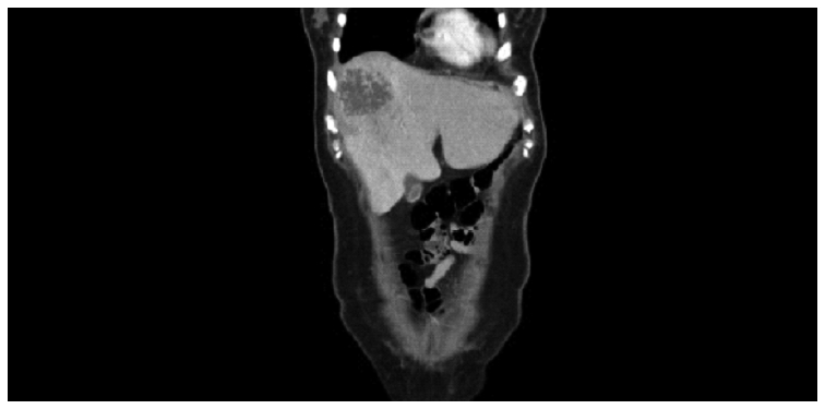
A 46-year-old female with a history of poorly controlled diabetes was admitted to the hospital with right upper quadrant abdominal pain, high fever, chills, and dyspnea lasting for recent 1 week. On admission, the patient’s body temperature was 38°C. Physical examination revealed severe tenderness in the right upper quadrant abdomen. Laboratory analyses produced the following results: 9.1 g/dL hemoglobin, 11,700/mm3 white blood cells count, 29 IU/L aspartate aminotransferase (AST), 90 IU/L alanine aminotransferase (ALT), 358 U/L alkaline phosphatase (ALP), 216 mg/dL blood glucose level, and 9.6% mg/dL hemoglobin A1c. A chest radiograph revealed a bilateral pleural effusion and patchy opacity in both lungs (Fig. 1A). An abdominal computed tomography (CT) scan showed a low-density lesion in segments 4 and 8. These findings led to the diagnosis of liver abscess, for which percutaneous drainage was performed. The cultured pus and blood samples were positive for K. pneumoniae resistant to ampicillin and ciprofloxacin, sensitive to ceftazidime and cefotaxime. The patient was treated with third-generation antibiotics (ceftriaxone) and metronidazole, but the clinical course did not improve. We thus discontinued these antibiotics and administered imipenem for 9 weeks. Follow-up imaging showed a reduction in liver abscess size and percutaneous drainage was removed (Fig. 2).

(A) A chest radiograph revealed a bilateral pleural effusion and patchy opacity in both lungs. (B) Chest computed tomography revealed multifocal areas of consolidation with ground-glass attenuation and a halo sign.

An abdominal computed tomography scan after percutaneous drainage removal showed the reduction in size of low-density lesion in segments 4 and 8.
An ophthalmic examination was performed because of the left eye vision difficulties. Endogenous endophthalmitis was diagnosed and an intravitreal injection containing 1mg vancomycin, 0.4 mg amikacin, and 2 mg ceftazidime was administered. However, the patient eventually lost her left eye vision completely.
Thoracentesis was performed and suggested exudation. A chest CT scan revealed multifocal areas of consolidation with ground-glass attenuation and a halo (Fig. 1B). These findings were compatible with pneumonia with septic lung due to K. pneumoniae -liver abscess.
With an appropriate antibiotic regimen, the patient’s clinical condition improved. There was no definite cause of pyogenic liver abscess. Colonoscopy was performed to investigate the underlying cause and revealed several polypoid lesions with shallow ulceration in the sigmoid colon and rectum (Fig. 3A). Polypectomy was performed by colonoscopy, revealing adenocarcinoma with submucosal second layer (SM II) (1,600 μm) invasion in one ulcerated polyp in sigmoid colon, and lamina propria in another polyp (Fig. 3B). The patient underwent an anterior resection of the colon. Histological examination of the surgical specimen revealed no residual malignancy, but metastasis was present in one of nine lymph nodes. The patient was diagnosed with staging III (T1N1M0) colon cancer and has been currently undergoing postoperative chemotherapy.
DISCUSSION
Liver abscess was associated with hepatobiliary tract disease or intra-abdominal infections, such as cholecystitis, suppurative cholangitis, appendicitis, diverticulitis, and peritonitis [1,2]. Some cases were identified as cryptogenic abscess. In several reports, some part of cryptogenic pyogenic liver abscess has been considered as the first manifestation of silent colorectal cancers and colonic tubulovillous adenoma, which bacteria may move from the mucosal defect in the colon into the portal system [4–6].
The most common bacteria isolated from liver abscess patients are gram-negative rods. Although Escherichia coli was the most commonly isolated from liver abscess patients before the 1980s, K. pneumoniae has recently been found to be the most common pathogen in Asia and is emerging in Western countries [7].
In patients with diabetics, liver abscess caused by K. pneumoniae is a known risk factor for embolic complications [8], especially endophthalmitis [9]. Septic metastatic lesions arising in liver abscess patients have been classified as endophthalmitis [9], septic pulmonary embolism [10], and chest wall abscess [2].
Septic pulmonary embolism is an uncommon but serious disorder that has been associated with certain risk factors, such as intravenous drug use, right-side bacterial endocarditis, pelvic thrombophlebitis, the presence of an indwelling catheter, and suppuration in the head or neck [11]. Infections by gram-negative organisms such as K. pneumoniae, which is the most common pathogen associated with liver abscess, may cause septic pulmonary embolism.
Endogenous endophthalmitis results from the hematogenous spread of the pathogen into the eye. This complication is due primarily to the high virulence of Klebsiella. Experimental animal models of intraocular infection have shown that the destruction of retinal photoreceptors begins as early as 24 to 48 hours after inoculation [11]. In our case, the patient lost vision in her left eye completely because she was admitted to the hospital 1 week after symptoms occurred.
The pathogenic mechanism of primary K. pneumoniae liver abscess complicated with metastatic infection remains unclear. Fang et al. [12] reported that mag A is an important virulence gene in invasive K. pneumoniae strains causing primary liver abscess and septic metastatic complications. The disruption of mag A results in complete loss of serum resistance and phagocytosis.
Pyogenic liver abscesses are considered cryptogenic when no primary source of infection is found. Cryptogenic cases account for one-third of liver abscesses [13]. Occult colonic cancer and tubulovillous adenomas have been recently reported in K. pneumoniae liver abscess patient [4,6]. In some cases, the causative relationship of underlying colorectal cancer was established only in retrospect, following clinical manifestation [5]. The presumed mechanism of liver invasion is the loss of integrity of the normal mucosal barrier allowing microbial seeding to portal venous system [13].
Lai and Lin [6] estimated the 5-year risk of colorectal cancer following a diagnosis of cryptogenic pyogenic liver abscess in 274 patients. They reported that the risk of colorectal cancer was 5.54 times higher in cryptogenic pyogenic liver abscess patients than in control patients. The odds of colorectal cancer were 4.08 times higher in females with cryptogenic pyogenic liver abscess than in control patients. These authors suggested that cryptogenic pyogenic liver abscess may signal colorectal cancer, especially in female patients with diabetes. They recommend that diabetic patients with cryptogenic pyogenic liver abscess undergo a colonic screening for the presence of a neoplasm.
Here, we reported a case of primary liver abscess caused by K. pneumoniae with multiple septic metastatic infections (endophthalmitis and septic pulmonary embolism) and colon cancer. Bacterial invasion to portal tract may have caused the pyogenic liver abscess. We recommended the genomic analysis of K. pneumonia, if possible, to aid in the prediction of the disease course. Although no formal guidelines currently recommended colonoscopy for liver abscess patients, in the setting of a “cryptogenic” pyogenic liver abscess, it seems prudent to include a thorough evaluation of the colon in the workup.
