Airway Responses to Deep Inspiration in Asthma by Different Bronchial Challenge
Article information
Abstract
Objective:
Deep inspirations (DI) provide physiologic protection against airway narrowing and DI-induced bronchoprotection and bronchodilation are impaired in asthma.
Methods:
To evaluate effect of DI on airway narrowing during methacholine challenge, we compared the 2 minutes tidal breathing method and the breath dosimeter method. Methacholine challenge in 12 asthmatics and 10 healthy controls was cross-overly performed by two methods. On first visit, a questionnaire for symptoms, allergy skin test, spirometry, and methacholine challenge was performed. On second visit, spirometry and methacholine challenge using the 25 mg/mL at 5 minutes intervals during the 2 minutes tidal breathing method and the ten-breath dosimeter method were performed on two separate days at same time each day.
Results:
The decreases in forced expiratory volume in one second (FEV1) and forced vital capacity during the 2 minutes tidal breathing method and dosimeter method in patients with asthmatics were higher than those in normal controls. The decreases in FEV1 and forced vital capacity during the 2 minutes tidal breathing method were higher than during dosimeter method in both asthmatics and controls.
Conclusion:
These observations indicate that the continuous generation method produce more bronchoconstriction than the dosimeter method during methacholine challenge and asthmatics had more bronchoconstriction than controls, suggesting inhibition of DI enhance methacholine induced airway narrowing in asthmatics.
INTRODUCTION
The pathophysiologic features of asthma include exaggerated airway narrowing to bronchoconstrictor agents and attenuated relaxation to β-adrenoreceptor stimulation. The abnormality of airway smooth muscle (ASM) is thought to be the airway hyperresponsiveness (AHR) that characterizes asthma [1]. The bronchodilatory effect of a deep inspiration (DI) and its failure in asthma, may lie glassy dynamics of the ASM cell [2].
AHR in asthma is believed to be caused in part by the inability of DIs to modulate airway narrowing. The ability to dilate methacholine-constricted airways by DI is impaired in subjects with rhinitis, possibly because of an abnormal behavior of ASM [3].
Lambert et al. [4] have attempted to determine how important an increase in smooth muscle mass could be as a contributor to exaggerated airway narrowing in a computer model of the tracheo-bronchial tree. They assumed that the contractile function and the length-tension relationship of asthmatic ASM was the same as normal, but that the force-generating capacity was increased in proportion to the increase in muscle mass. The force per cross-sectional area (stress) was significantly greater for asthmatic subjects in studies evaluating the cross-sectional ASM in asthmatic versus control preparation [5]. Asthmatic preparations produce a two-to threefold increase in shortening of smooth muscle compared with controls, suggesting that increased ASM shortening is a major determinant of the excessive luminal narrowing seen in asthma [6]. Inflammation in the ASM bundles and submucosa of bronchial biopsies is positively associated with impaired airway mechanics during DI in asthma [7]. DI regulates airway responsiveness at the airway level, but this is limited by airway stiffness due to reduced ASM strain in animal model [8].
Bronchial inhalation challenge tests have been used for several decades as means of modeling asthma for investigative purposes. Bronchial challenge tests may be used to assess airway reactions to specific allergens and other sensitizing agents and also to quantify non-allergic airway responsiveness to pharmacological agents such as methacholine or histamine. Two particular methods of aerosol delivery such as the intermittent generation of aerosols during inspiration and continuous aerosol generation during tidal breathing are commonly used and give remarkably similar result [9]. Prohibition of DI during methacholine challenge produces more bronchoconstriction than a conventional challenge, which mandates deep breaths in order to perform forced expiratory volume in one second (FEV1) measurement [10,11]. A submaximal inhalation dosimeter methacholine challenge results in a significantly lower PC20 compared with the standard 5-breath dosimeter method, presumably because of the bronchoprotective effect of a DI required for the latter [12].
In the present study, we compared the FEV1 and forced vital capacity (FVC) during the 2 minutes tidal breathing method and during the ten-breath dosimeter method to assess differences of airway narrowing during methacholine challenge in asthmatics and normal controls.
MATERIALS AND METHODS
1. Subjects
Twelve asthmatics and ten subjects were participated in this study (Table 1). We enrolled patients who presented to the Division of Allergy and Respiratory Medicine in Soonchunhyang University Hospital because of asthma, defined as one or more asthma symptoms, and whose physical examinations were compatible with the American Thoracic Society’s definition of asthma [13]. Each patient showed airflow reversibility, as documented by a positive bronchodilator response with a greater than 15% increase in FEV1 and/or airway hyper-reactivity to less than 10 mg/mL methacholine [14]. And each patient had good control status of asthma during experiments period with inhaled steroid. The exclusion criteria included greater than 10-pack years smoking, less than 20 or more than 70 years old, parenchymal lung disease apparent on chest radiography, diffusing capacity less than 80%, severe uncontrolled diabetes mellitus, and renal, hepatic, or cardiac failure. This study was performed with the approval of the ethics committee of the Soonchunhyang University Hospital, and informed written consent was obtained from all of the study subjects.
The control subjects who volunteered for this study had no history of any respiratory symptoms, and had FEV1 >75% of predicted value, and had a ratio of FEV1 to FVC >75%. No subject had any respiratory infections for 4 weeks prior to the study. Coffee or tea was not allowed before the tests on the day of the test.
2. Study design
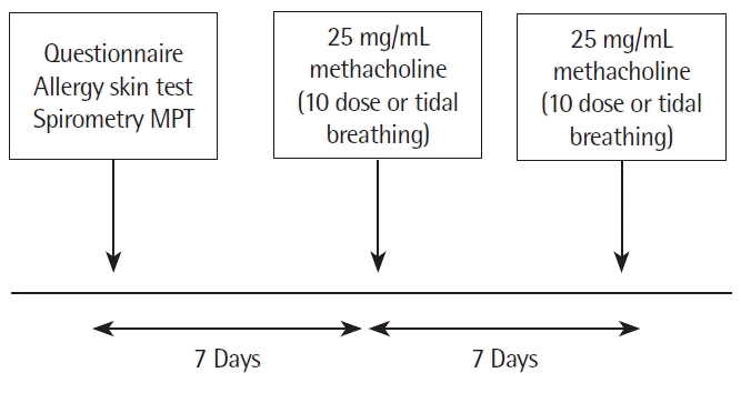
On first visit, a questionnaire for symptoms, allergy skin test, spirometry, and methacholine challenge testing (Fig. 1) were performed. Spirometry was performed with Multispiro spirometer (SX/PC; Creative Biomedics, San Clemente, CA, USA). Complete flow-volume curves were generated, and FEV1 and FVC were measured. Measurements were accepted if two or more values of FEV1 and FVC were within 5% of each other, and the mean of these values was used at the baseline value. All values were recorded in liters at body temperature-atmospheric pressure-saturated, and were expressed as percents of predicted values based on the equations of Crapo et al. [15].

Study protocols. Methacholine provocation tests (MPT, conventional 2 minutes tidal breathing and five-breath dosimeter method) were cross-overly performed at seven days interval. Subjects cross-overly underwent 25 mg/mL methacholine challenge (2 minutes tidal breathing and ten-breath dosimeter method) on two separate days at same time each day).
At first visit methacholine challenge tests (2 minutes tidal breathing) was carried out by some modifications of the methods according to guideline for methacholine and exercise challenge [13]. And then methacholine challenge tests were cross-overly carried out by some modifications of the methods according to guideline for methacholine and exercise challenge [13] (2 minutes tidal breathing and ten-breath dosimeter) on two separate days at same time each day. Allergy skin prick tests with commercially available inhalant allergens (Dermatophagoides pteronyssinus, Dermatophagoides farinae, Aspergillus spp, alder, birch, hazel, rye, timothy, mugwort, ragweed pollen, cockroach; Allergopharma Co., Hamburg, Germany) and histamine (1 mg/mL; Histamine, Allergopharma Co.) were performed on the volar side of both forearms. After 15 minutes, the mean diameter of a wheal formed by the allergen was compared with that formed by histamine. If the former was the same or larger than the latter (allergen/histamine [A/H] ratio≥1.0), it was considered positive. Atopy was defined as a reactor who showed A/H ratio≥1.0 to one or more allergens on skin prick tests [16].
On second visit, five doses of methacholine (25 mg/mL) challenge tests were cross-overly performed by two methods (ten-breath dosimeter and 2 minutes tidal breathing) on two separate days at same time each day. In brief, by dilution in buffered saline (pH 7.4), methacholine (Sigma Chemical, St. Louis, MO, USA) at concentrations of 25 mg/mL was prepared. On first day five 25 mg/mL doses of methacholine were given every 5 minutes by Pari Boy nebulizer (PARI GmbH, Starnberg, Germany), for 2 minutes, and spirometry was conducted 1 minute after each dose. On subsequent day, A Micro dosimeter (S&M Instrument Co., Doylestown, PA, USA), triggered by a solenoid valve set to remain open for 0.6 seconds, was used to deliver the aerosol generated from a DeVilbiss 646 nebulizer with pressurized air at 20 psi. Each subject inhaled 10 inspiratory capacity breaths of buffered saline and 25 mg/mL doses of methacholine every 5 minutes, and spirometer was conducted 1 minute after each dose. FEV1 was measured with a computerized spirometer (SX/PC; Creative Biomedics) at 1 minute after each inhalation was adopted for analysis. The recovery time from maximal decrease in FEV1 and FVC to baseline FEV1 and FVC was checked without using bronchodilator after methacholine challenge test.
3. Statistical analysis
All data were analyzed using the SPSS ver. 10.0 (SPSS Inc., Chicago, IL, USA). Data are expressed as mean±standard error of mean. Comparison of the decrease in FEV1 and FVC on the 2 minutes tidal breathing method with the decrease in FEV1 and FVC on the ten-breath dosimeter method after the same dose methacholine was performed using paired t-test for variables. A P-value of < 0.05 was considered significant.
RESULTS
The characteristics of subjects are given in Table 1. Ten control subjects and 12 asthmatics went to the five doses of 25 mg/mL methacholine during the ten-breath dosimeter and the 2 minutes tidal breathing method. One of control subjects and two of asthmatics was stopped after two doses of 25 mg/mL methacholine during the 2 minutes tidal breathing method because of large decrease in FEV1 (of 72.5% of baseline) and shortness of breath. But the control subjects and asthmatics went to the five doses of 25 mg/mL methacholine during the ten-breath dosimeter method.
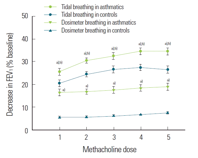
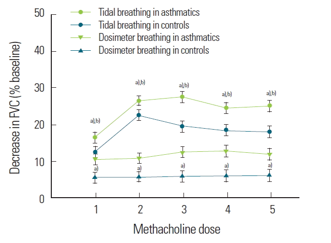
The mean decreases in FEV1 and FVC during the 2 minutes tidal breathing method were 31.8%±4.3% and 24.4%±5.1% in asthmatics and 22.2%±5.3% and 16.8%±5.1% in normal controls, respectively. The mean decreases in FEV1 and FVC during the ten-breath dosimeter method were 12.2%±5.1% and 10.8%±4.9% in asthmatics and 8.0%±2.3% and 5.2%±3.1% in normal controls. The FEV1 at methacholine dose 1, 2, 3, 4, and 5 were significantly greater decreased during the 2 minutes tidal breathing method compared to those during the ten-breath dosimeter method (Fig. 2). The FVC at methacholine dose 1, 2, 3, 4, and 5 were significantly greater decreased during the 2 minutes tidal breathing method compared to those during the ten-breath dosimeter method (Fig. 3). The dyspnea perception according to Borg score at methacholine dose 1, 2, 3, 4, and 5 were significantly greater decreased during the 2 minutes tidal breathing method compared to those during the ten-breath dosimeter method in asthmatics and normal controls (Fig. 4).

Decrease in forced expiratory volume in one second (FEV1) between the 2 minutes tidal breathing method (upper squares) and the ten-breath dosimeter method (lower circles) during methacholine challenge. a)P< 0.05 compared to controls. b)P< 0.01 compared to dosimeter breathing methods.

Decrease in forced vital capacity (FVC) between the 2 minutes tidal breathing method (upper squares) and the five-breath dosimeter method (lower circles) during methacholine challenge. a)P< 0.05 compared to controls. b)P< 0.01 compared to dosimeter breathing methods.
DISCUSSION
The results of this study indicate that the continuous generation method produce more bronchoconstriction than the dosimeter method for delivering methacholine aerosol during methacholine challenge test and asthmatics had more bronchoconstriction than controls, suggesting inhibition of DI enhance methacholine induced airway narrowing in asthmatics.
The principal features of asthmatic airways include exaggerated narrowing to bronchoconstrictor agonists and attenuated relaxation to β-adrenoreceptor stimulation. These functional alterations are associated with inflammation and remodeling of the airways, which include an increase in ASM cell mass, disruption of the airway epithelium, and changes in the airway tissue extracellular matrix [1]. Increased thickness of the airway may have an important amplifying effect on the contractile response of ASM and may be a major mechanism contributing to AHR in asthma [16]. Increased ASM mass, which may be induced by myocyte hypertrophy or hyperplasia, has been a well-recognized pathologic finding in the bronchi of patients with chronic severe asthma. The increase in ASM cell mass, coupled with inflammation-induced changes in the surrounding extracellular matrix, may reduce the load acting on the muscle due to peribronchial inflammation and give rise to enhanced ASM contractility [17,18].
Bronchoconstriction can occur due to direct action on ASM cells by methacholine, histamine, and cysteinyl leukotrienes and indirect action by releasing bronchoconstrictors from inflammatory cells and nerves [19,20]. The distinguishing feature of asthma was a failure of a DI to reverse airway narrowing rather than an exaggerated capacity of the airways to narrow [21]. Airway hyperactivity is perhaps not so much a stimulus as a reflection of an impaired ability to modify changes in bronchomotor tone by lung inflation [21]. In comparison of the dosimeter and the continuous generation method for delivering histamine aerosol, the efficiency of airway delivery of aerosol (lung dose as a percentage of dose delivered to the mouth) was 26% for the dosimeter system compared to 4% for Wright nebulizer system [9]. Airway closure during induced bronchoconstriction is more resistant to the effects of DI in asthmatic than in normal subjects but the reversibility of bronchoconstriction by DI is not different [22]. In the present study, the authors evaluated effects of different method used commonly during methacholine challenge on airway narrowing. We compared the airway response to methacholine using 2 minutes tidal breathing inhalations of the 25 mg/mL concentration of methacholine at 5 minutes intervals, with the counted breaths technique looking at ten inspiratory capacity breaths repeated on five occasions at 5 minutes intervals again using 25 mg/mL dose. The FEV1 and FVC in asthmatics and normal controls were significantly greater decreased during the 2 minutes tidal breathing method compared to those during the five-breath dosimeter method, suggesting inhibition of DI may enhance methacholine induced airway narrowing in normal control and asthmatics, suggesting that DI inhibition have more bronchoconstriction in both asthmatics and normal control in consistent with study of Skloot et al. [23] reported that DI protects against methacholine-induced bronchoconstriction in healthy subjects and a fast DI is bronchoprotective while a slow DI is not. Also bronchoconstriction during methacholine challenge was more higher in asthmatics than in normal controls and tidal breathing method more decrease in FEV1 and FVC than dosimeter method in contrast with that the magnitude of DI-induced bronchodilation is only slightly reduced in mild asthmatics, compared to healthy subjects, supporting to the hypothesis that the attenuation of the bronchodilatory effect of DI contributes to the severity of the clinical manifestations of asthma [24].
Skloot et al. [25] suggested that prohibition of DI in normal subjects make them resemble asthmatics in that bronchodilation after DI is reduced. Prolonged prohibition of DI may allow ASM to become stiff and noncompliant and/or lead to a failure of transmission of the stretch provided to the muscle by DI. The effect of a DI may be larger in peripheral airways than in central airways. The structures in membranous bronchiloli are subjected to traction by surrounding tissues, and a maximal inspiration may cause a larger distending force in the peripheral airways than in larger bronchi [26].
The DI associated with the FEV1 maneuver decreases the bronchial tone in airways constricted by methacholine for up to 6 minutes, possibly due to yielding of cross links in ASM [27]. King et al. [28] suggested that there be several possible contributory mechanisms for the effects of serial DI maneuver in contracted airways, such as changes in lung volumes, elastic fibers, surfactant, or release of mediators. They reported that the increased airway narrowing associated with inhibition of DI during ASM contraction occurred after 10 minutes in normal subjects, at which time the response plateaus. In this study the more decrease in FEV1 is, the longer recovery time to base line FEV1 is, indicating that the increased airway narrowing during methacholine challenge is associated with recovery time of ASM contraction.
Bronchodilation following DI is usually impaired in patients with asthma. This might be due to changes in airway mechanics in the presence of inflammation or structural changes within the airways. Systemic antiinflammatory treatment in addition to maintenance therapy with inhaled corticosteroids increases bronchodilation by DI in patients with mild-to-moderate persistent asthma. This suggests that residual inflammation impairs airway mechanics in asthma patients [29].
DI-induced bronchoprotection and bronchodilation are impaired in asthma. DI-induced bronchoprotection can be restored by inhaled glucocorticosteroids only in individuals with mild hyper-responsiveness. After steroid treatment, the beneficial effects of DI become significant determinants of the magnitude of AHR [30]. But it should be considering that both methods provide similar values for slope and level of plateau, suggesting that the technical factors that affect methacholine sensitivity and the shape of the curve are different [31].
In conclusion, this study shows that the continuous generation metho enhance methacholine induced airway narrowing in normal controls and asthmatics and more methacholine induced airway narrowing in asthmatics indicating that inhibition of DI enhance methacholine induced airway narrowing in asthmatics.
Acknowledgements
This study was supported by the grants of the Korea Health 21 R&D Project Ministry for Health, Welfare and Family Affairs, Korea, A102065).

