INTRODUCTION
Rosai-Dorfman disease (RDD) is a proliferative lesion of histiocytes, predominately affecting children and young adults. Most patients with this disease are presented with bilateral cervical painless lymphadenopathy. Most often, extranodal manifestations of the disease are soft tissue of the head and neck, paranasal sinuses [1]. Histological hallmark of RDD is emperipolesis, which refers to the presence of variable numbers of intact lymphocytes within the cytoplasm of histiocytes. It usually has a self-limiting process, but can rarely be fatal when systemic involvement happens. There are several treatment options, including steroid, chemotherapy, radiotherapy, and surgical debulking according to the symptoms or the extent and localization of the lesions. We report a very rare case of cutaneous RDD combined with aortic vasculitis, arrhythmia and valvular heart disease, which were treated with prednisolone and methotrexate (MTX) showing modest improvement of vasculitis.
CASE REPORT
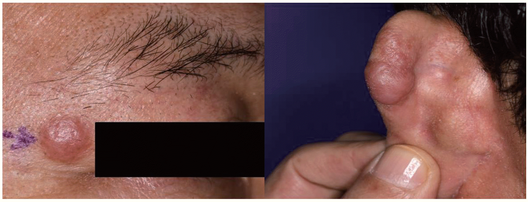
A 54-year-old man was hospitalized to evaluate chest tightness, epigastric pain, diaphoresis, which began 3 days ago, and facial skin lesions, of which, size had been slowly increased for 2 months. Physical examination showed no structural abnormality around the chest with clear breathing sound and regular heart beat. Three nodular skin lesions were found at the right eyelid and both auricles without itching sense, pain and tenderness (Fig. 1).
The initial laboratory investigation showed a white blood cell count of 9,930/μL, hemoglobin level of 11.4 g/dL, and platelet count of 362×103/μL. The lactate dehydrogenase level was 169 IU/L (normal range, 106 to 211 IU/L); C-reactive protein was elevated to 5.1 mg/dL; and erythrocyte sedimentation rate, to 113 mm/hr. Autoimmune antibodies, including anti-nuclear antibody, anti-citrullinated protein antibody, rheumatoid factor, and anti-neutrophil cytoplasmic antibody, were negative. Bone scan showed no abnormal increased bony uptake.
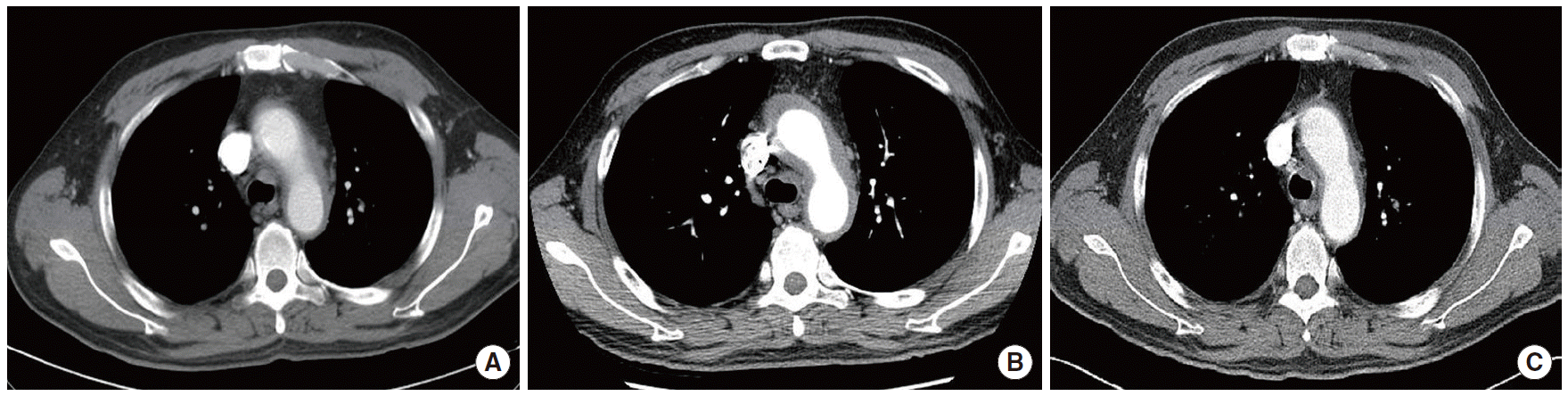
Transthoracic echocardiography (TTE) revealed no specific abnormal findings with preserved left ventricular function and normal valvular movement. A computed tomography (CT) scan showed circumferential wall thickening in the ascending aorta, aortic arch and proximal descending thoracic aorta, implicating vasculitis (Fig. 2A). Even though a number of causes for aortic vasculitis are possible, such as syphilis, autoimmune vasculitis, giant cell arteritis, Takayasu’s arteritis, and rheumatoid arthritis, without biopsy from affected aorta and relevant autoimmune antibodies from laboratory tests, we could not get final diagnosis on aortic vasculitis.
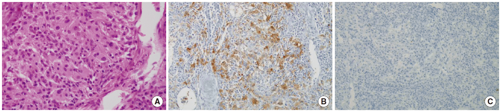
The biopsy on skin lesion of the right auricle was performed and its’ result was consistent with cutaneous RDD disease showing a massive infiltration of histiocyte with lymphophagocytosis, and positive for S-100, but not CD1a (Fig. 3A-C). We decided to closely observe without any treatment. After 2 months, he complained newly developed palpitation, and the size of the skin lesions was increased. Holter monitoring indicated premature ventricular complexes, which did not require active treatment. Thoracic and abdominal aortic wall thickening and pulmonary arterial wall thickening in a CT scan were slightly progressed (Fig. 2B), and mild tricuspid and mitral valvular regurgitation was also newly observed in TTE.
Therefore, we started electron radiotherapy of the total dose of 10 Gy (five times of 2 Gy) to skin lesions and oral prednisolone (2 mg/kg/day) for two weeks. The frequency of palpitation decreased, but skin lesions were not changed. Three times of intravenous MTX (each 15 mg) had been challenged every others day for six days, which was stopped for liver enzyme elevation without any changes of skin lesions and valvular regurgitation. Follow-up of CT scan 2 month later showed the slight improvement of vasculitis after these treatments (Fig. 2C). To prevent the disease progression, continuous hydroxychloroquine (400 mg/day) has been administered for more than two years and the symptom and sign are maintained as the stationary state so far.
DISCUSSION
RDD is a rare and benign self-limited disorder that was described by Rosai and Dorfman in 1969 [1]. The most common involving site is the lymph node (87%), followed by the skin/soft tissue and nasal cavity/paranasal sinuses. The other involving sites are the orbits, eyelids, salivary glands, heart, upper respiratory tract, bone, kidney, and central nervous system. The main presenting symptoms depend on the involved sites, such as swollen neck mass, tonsillitis, nasal discharge, and obstruction [2]. Histologically, the lymph node sinuses expanded by a proliferation of distinctive histiocytes are seen, and the hallmark of RDD is the presence of variable numbers of intact lymphocytes within the cytoplasm of histiocytes, a phenomenon referred to as emperipolesis. The immunohistologic studies for RDD reveal the expression of the S100 protein and negative for CD1a, and variably positive for CD68. One of the most important differential diagnoses from RDD is the Langerhans cell histiocytosis, which shows positive for both the S100 protein and CD1a [3].
The cases with skin involvement of RDD have been reported from purely skin involvement without lymphadenopathy to a form combined with the extranodal organ [4,5]. RDD with pathologically vasculitic feature was reported in the skin lesions of the petechiae and palpable purpura [6]. In our case, vasculitis was found in great vessels of the aorta and pulmonary artery, but not in the skin lesion. These vasculitic lesions were slightly improved after steroid and MTX treatment.
There are a few reports on extranodal involvement because of rarity of RDD itself, with even fewer reporting on cardiac involvement. Yontz et al. [7] reported one interesting case of RDD, which involved at heart as mass feature confirmed by a needle biopsy. In our case, this rare cardiac involvement of RDD presented as vasculitis of great vessels, frequent premature ventricular complex and valvular dysfunction.
There are several treatment options for RDD according to the symptoms or the extent and localization of the lesions. If the RDD is not massive and do not involve the vital organs, only observation is recommended generally, and many physicians have reported complete remission of RDD without any treatment [8]. Vital organ involvement or compression resulting in life- threatening manifestations should be treated by surgical debulking and/or radiotherapy [9]. While the efficacy of chemotherapy is still in controversy, combination chemotherapy with or without prednisone has shown the effect to RDD, even complete remission [10]. In our case, the patient has been treated with radiotherapy to the skin lesion, which had no effect on it. However, oral prednisolone (2 mg/kg/day) for 2 weeks and intravenous MTX (three times of 15 mg) led modest improvement of the symptoms of heart involvement and vasculitis of the great vessels.
In conclusion, we reported a very rare case of RDD involving the facial skin with unusual systemic manifestation, including vasculitis of the great vessels, arrhythmia, and valvular dysfunction, which were treated with steroid and MTX resulting modest improvement of vasculitis.