INTRODUCTION
Dacryocele is an uncommon congenital condition of nasolacrimal drainage system with a grey bluish mass like appearance on the medial canthus, which is a result of nasolacrimal duct obstruction, both proximal and distal [1]. The pathogenesis of the obstruction is the result of an upper obstruction of Rosenmuller valve and lower obstruction of the Hasner valve [2]. This mechanism can lead to accumulation of fluid in lacrimal sac, resulting secondary infection, which may require a prompt treatment for the obstruction of drainage system. Other complication includes intranasal cysts, astigmatism induced by compression [3].
Most frequently used imaging modality is ultrasound (US), which is a simple and non-invasive way of distinguishing dacryocele from other similar pathologic conditions [4], and other imaging modalities, such as computed tomography (CT) or magnetic resonance imaging (MRI), can also help diagnosing dacryocele [5,6]. In this article, we present a case of bilateral dacryocele which showed typical findings of dacryocele in orbit MRI images.
CASE REPORT
Otherwise healthy, full term, 46-day-old boy was referred for evaluation of bilateral orbital mass on both medial canthus, with yellowish eye waxes which started since 7 days of age. The parents have visited local hospital and were instructed to compress the lacrimal sac area frequently, but the mass did not resolve.
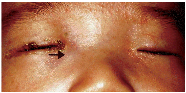
On external examination, the palpable bluish masses were seen on both medial canthus, with eye waxes more prominent on his right eye. The patient was on intravenous antibiotics and topical tobramycin eyedrop, and had taken orbit MRI for differential diagnosis, to rule out other orbit and eye anomalies.
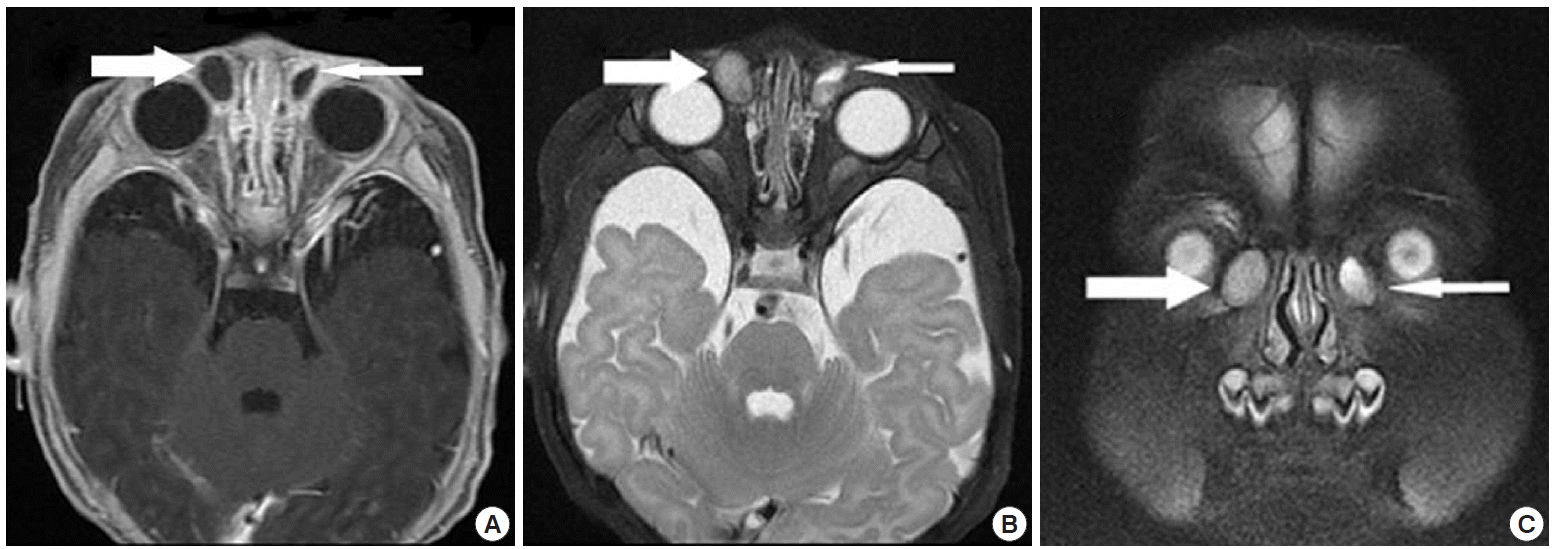
On orbit MRI images with enhancement, we could easily observe well encapsulated lacrimal sac mass in his both nasolacrimal duct, with low signal on T1- and high signal intensity on T2 weighted images, and the size of lesions were 11.5×6.5 mm for right, and 11.5 ×8.5 mm for left (Fig. 1). Fluid level was visible on the left side lesion (Fig. 1A, B). Since MRI findings did not show any sign of inflammation, all antibiotic treatments were stopped, and after 3 days without any further management, the mass on the left had spontaneously resolved and the size was decreased on the right (Fig. 2).
DISCUSSION
A dacryocele usually presents as an enlarged blue cystic lacrimal sac at birth [1]. The nasolacrimal drainage system develops from surface ectoderm located in the nasal and maxillary process, and canalization of the drainage system starts at week 9 [7]. Hasner valve level is the last barrier to canalization [7]. It is widely accepted that a dacryocele is developed from the persistent membrane at the level of this barrier and obstruction of Rosenmuller valve [2]. Fluid accumulation occurs, and secondary infection may develop.
It is important to distinguish dacryocele from other similar pathologic conditions, because initial treatment can be very different. The pathologic conditions which can mimic dacryoceles include hemangioma, encephalocele, glioma, dermoid cysts, and malignant, and may need further imaging evaluation [8]. Also, early detection of any complication is necessary to manage patients properly. Dacryocele without secondary infection requires only conservative approach (i.e., warm compress, massage), but when it comes to complication, such as dacryocystitis and cellulitis, probing and antibiotic treatment may require [9].
Commonly, dacryocele is diagnosed with US for its safe characteristics: non-invasive and radiation free. High-resolution US is widely used for detecting any pathologic orbit and eye condition, especially in prenatal period, and is also best chosen for differential diagnosis. A medial cystic mass, in communication with the dilated nasolacrimal duct, in addition to the fluid and debris content, is a typical US finding of dacryocele. US may be useful in differential diagnosing, since high internal reflectivity is observed in other mimicking condition such as dermoid cyst and hemangioma. CT and MRI are also used for diagnosing such abnormalities involving the orbit and eye, but commonly used as secondary modality next to US. CT has the advantage of detecting any calcification and bone changes, which may be observed in dermoid cysts, but not in dacryoceles, whereas MRI has the advantage of presenting soft tissue changes, such as cystic changes, without any radiation exposure [8]. MRI can provide evidence for differentiating dacryocele from other pathologic condition with similar appearance by showing anatomical continuity of a medial canthal cystic mass with enlarged nasolacrimal duct [10]. Since CT and MRI are conducted only when the diagnosis is inconclusive with US finding, there are only limited numbers of articles on CT and MRI findings of dacryocele, with literatures on MRI descriptions especially lacking.
In this article, we found MRI can be useful in delineating the lesion. Also, MRI findings can aid in characterizing the content of the lesion. From the low signal on T1- and high signal on T2-weighted images, we could predict that the cystic mass was filled with mucoid material. With such advantages, MRI can be useful imaging modality to define the margin of the lesion and the characteristics of inner contents, aiding in differential diagnosis from other similar anomalies. Thus, this article may provide valuable information on orbit MRI findings of dacryocele to other clinicians those interested in congenital nasolacrimal system anomalies.


