INTRODUCTION
Clinical manifestation of hepatotoxicity of herbal medication is various and ranged from symptoms of self-limiting hepatitis to fulminant hepatitis that result liver transplantation or death. The diagnosis of Wilson’s disease based on clinical findings, biochemical tests, liver biopsy, and genetic studies has been reviewed recently and diagnostic algorithms using critical parameters have been suggested [1]. Sometimes, the exact diagnosis might not be able to be established based on this criteria only, thus caution is essential for making an accurate diagnosis of Asian patients with history of herbal medication.
CASE REPORT
A 39-year-old female, who was overweighted (body mass index, 29.12 kg/m2) was admitted to Kyung Hee University Hospital at Gangdong with 2 weeks of vague abdominal discomfort, anorexia, nausea, dyspepsia, fatigue, and jaundice. In an effort to lose weight, she had been taking herbal powder which is natural supplement for weight management containing hydroxycitric acid, three times a day for 2 days, from 12 days prior to admission up until the development of the symptoms. She was not a heavy drinker of alcohol, drinking a bottle of beer per week. She did not take any prescribed medications. She denied taking any other supplements or alternative medications except the herbal powder. Herbal power that the patient took included aloe powder, kelp powder, stevioside, extract of green tea, powder of prune, powder of cereal enzyme and extracts of Garcinia cambogia and the main ingredient of herbal powder is extracts of Garcinia cambogia for weight loss.
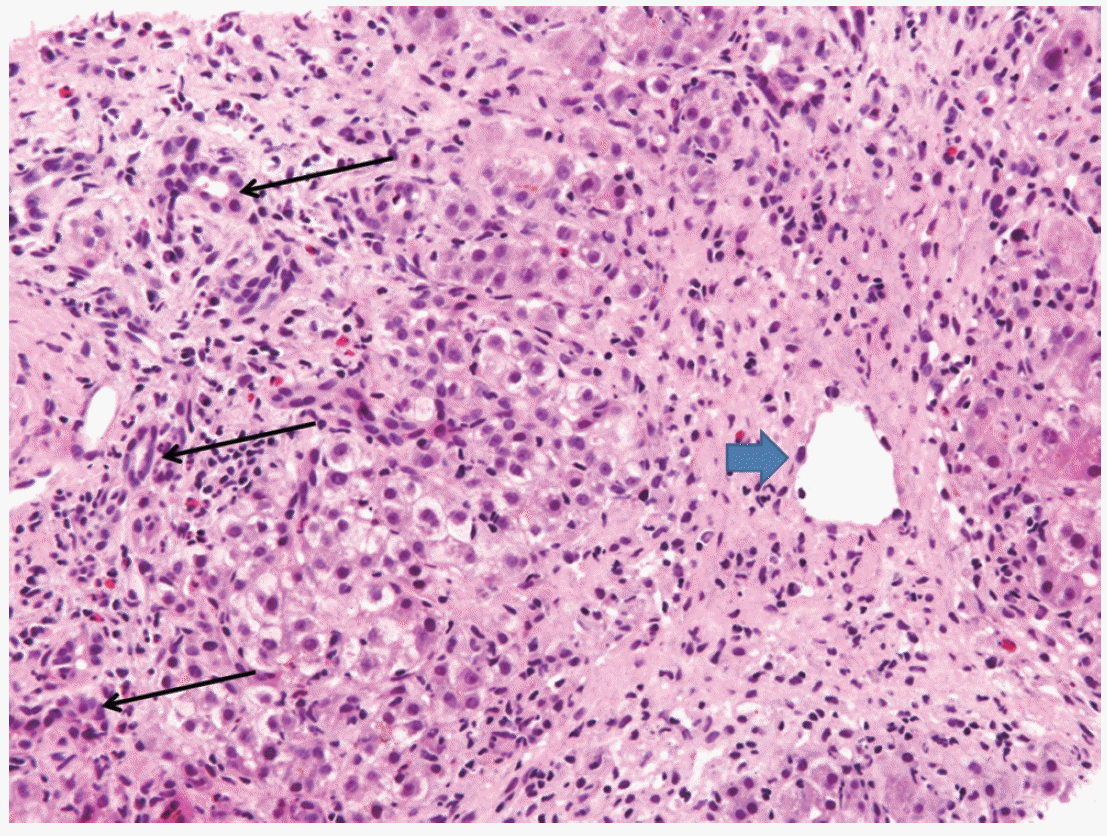
She was afebrile with stable vital signs. Physical examination was remarkable for jaundice and icteric sclerae. She had no liver enlargement and no stigmata of chronic liver disease. Laboratory analysis revealed a serum aspartate aminotransferase (AST) of 332 U/L, alanine aminotransferase (ALT) of 541 U/L, alkaline phosphatase of 324 U/L, total bilirubin (TB) of 10.9 mg/dL, albumin level of 3.2 g/dL, prothrombin time of 22.2 seconds (international normalized ratio of 2.03), normal complete blood count, normal electrolyte panel, and normal estimated glomerular filtration rate. Diagnostic evaluation was negative for hepatitis A, B, C, cytomegalovirus, EpsteinBarr virus and autoimmune liver disorders such as anti-mitochondrial antibody, anti-liver/kidney microsomal antibody and antismooth muscle antibody. Serum copper level was 87 μg/dL (range, 70 to 130 μg/dL) and ceruloplasmin level was decreased to 15 mg/dL (range, 20 to 40 mg/dL). Twenty-four-hour urine copper level was not increased to 30 μg/dL (range, 38 to 70 μg/dL). Slit-lamp examination for Kaiser-Fleischer rings was negative. A computerized tomography scan of her abdomen showed no abnormal attenuation of the liver. However, her liver biopsy specimens revealed lobular necrosis, fibrosis and cholestasis suggestive of drug-induced liver injury (Fig. 1). Quantitative hepatic copper was 204 μg/g dry weight (range, 10 to 35 μg/g dry weight). The decreased serum ceruloplasmin and increased hepatic copper level supported the diagnosis of Wilson’s disease but the increment of liver copper level did not reach diagnostic level [1]. Thus, following the diagnostic algorithm proposed by Roberts et al. [1], mutation analysis by whole-ATP7B gene sequencing was performed, and no mutations were identified including R778L, A874V, and N1270S known as the three most common mutations associated with Wilson Disease in a Korean population [2]. When putting together the above mentioned contents, the Councils for International Organizations of Medical Sciences/Roussel Uclaf Causality Assesment Method scale come out be 9 points, which is appropriate to highly probable (score>8 points) casuality of drug induced liver injury.
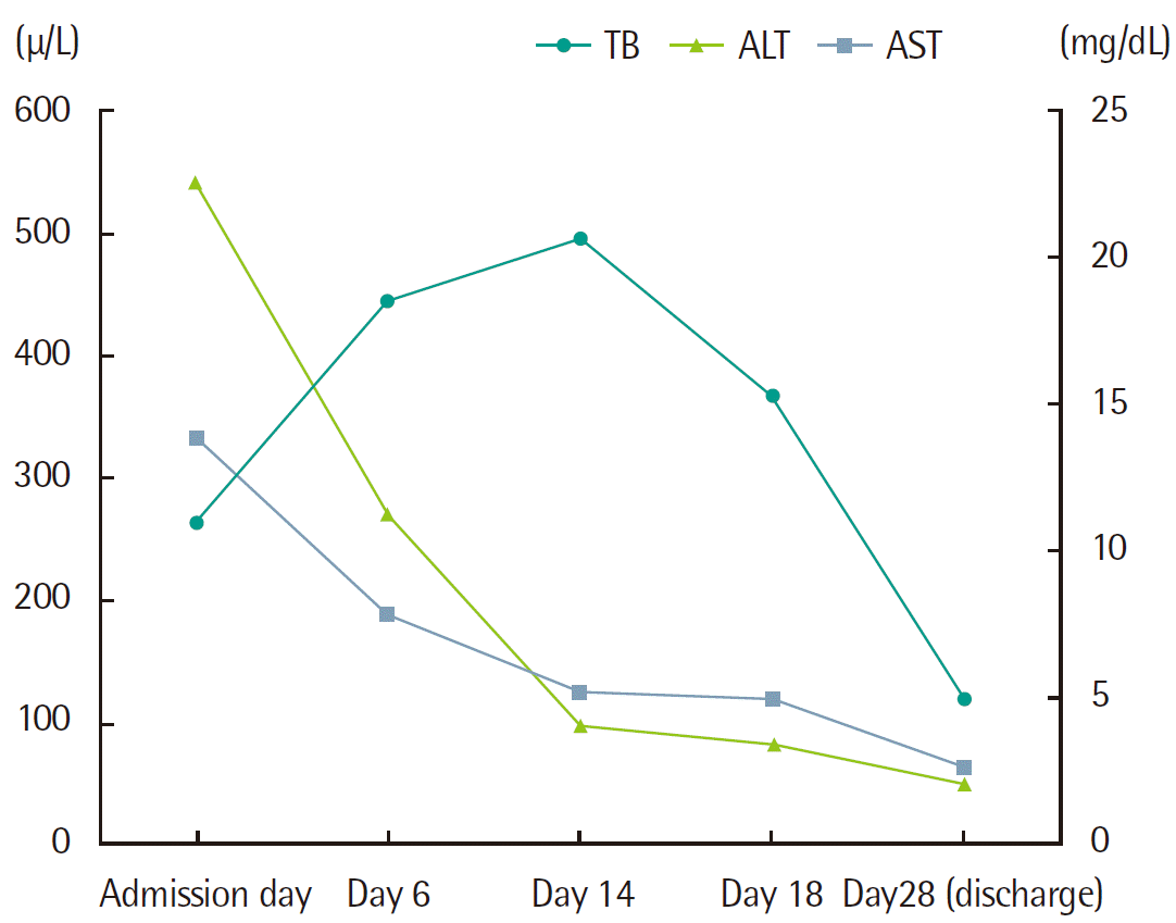
She was treated supportively with fluid and nutrition. Her liver function tests gradually normalized from the day of admission except for TB. The bilirubin level peaked on hospital day 14 with serum TB level of 20.6 mg/dL and direct bilirubin level of 16.1 mg/dL. On hospital day 28 (day of discharge) her liver function tests showed a AST level of 64 U/L, ALT level of 50 U/L, and TB of 5.0 mg/dL (Fig. 2). The patient did not represent coagulopathy, hepatic encephalopathy or any other complications. She was followed up for four months and her symptom and sign such as icteric sclera, jaundice, and dyspepsia continued to improve steadily. Four months later, she visited out patient clinic. Her pruritus and jaundice had resolved completely. The patient’s liver test showed a serum AST level of 37 U/L, serum ALT level of 41 U/L with her good condition.
DISCUSSION
Clinical presentations of toxic hepatitis are variable, so it is not possible to make a clear cut diagnosis as ‘toxic hepatitis’ only by history of a medicine use. Thus differential diagnosis of toxic hepatitis is essential to rule out other liver disease and make a correct diagnosis. Laboratory tests for viral hepatitis, auto-immune disorders and various metabolic diseases should be performed in this context. The test results of the patient with toxic hepatitis can mimic other disease. In terms of our case, even if declined serum ceruloplasmin and increased hepatic copper level implied the possibility of underlying Wilson’s disease, other diagnostic parameters such as 24-hour urine copper level, family history of Wilson’s disease, neurological manifestation, and Kaiser-Fleischer rings on slit lamp examination failed to meet the criteria for Wilson’s disease.
A low serum ceruloplasmin level may make physicians consider the diagnosis of Wilson’s disease. Ceruloplasmin is a copper-carrying protein that is bound to 90% of the circulating copper in normal individuals. The reference range of serum ceruloplasmin level is 20 to 40 mg/dL, and the concentration below 20 mg/dL is suggestive of Wilson disease [3]. However, the ceruloplasmin level has limited practical use, because the ceruloplasmin concentrations under 20 mg/dL is able to be found in 1% of controls, in 10% of heterozygous Wilson disease carriers and in patients with copper deficiency, Menkes disease, hereditary hypoceruloplasminemia, malabsorption, nephrotic syndrome, and chronic liver failure [4-8]. Under normal physiological conditions about 98% of the copper excretion is via the bile and only 2% via the urine [9]. Accordingly, in the case of the bile flow interruption such as cholestasis, copper comes to be accumulated in the liver [10]. In short, hepatic copper concentration is expected to be elevated in cholestatic condition such as biliary atresia or other situation of hepatic congestion, since bile ducts are blocked and cholestasis persists [9]. In 1995, Bayliss et al. [11] reported that hepatic copper concentration was high in surgical specimens of the biliary atresia patients, though the hepatic zinc and manganese level were low. Hepatic copper content >250 ug/g dry weight remains the best biochemical evidence for Wilson’ s disease [1,12]. Our patient’s declined ceruloplasmin level might be due to her mal-nutrition in her effort of weight reduction. Her increased liver copper level could be attributable to cholestasis consistent with hepatotoxicity associated with dietary herbal supplement.
The manufacturer’s list of ingredients in the dietary herbal supplement is dietary fiber, aloe powder, kelp powder, stevioside, extract of green tea, powder of prune, powder of cereal enzyme and extracts of Garcinia cambogia, etc. The exact portion of the contents is not demonstrated in the list. Extracts of Garcinia cambogia have been associated with cases of severe hepatotoxicity among those ingredients. Stevens et al. [13] reported on cases of two patients with acute liver injury associated with the herbal weight-loss supplement, hydroxycut, which contains potentially hepatotoxic hydroxycitric acid (HCA). HCA comes from the Garcinia cambogia, tropical fruit. Fong et al. [14] analyzed a case series of 17 patients with hepatotoxicity due to hydroxycut. In this report, the authors sought to characterize the clinical presentation of hydroxycut-associated liver injury and reported that usual symptoms were jaundice, fatigue, nausea, vomiting, and abdominal pain. Most patients exhibited a hepatocellular pattern of injury and three required liver transplantation [14]. Our patients presented with jaundice, fatigue, abdominal discomfort also despite the small amount of ingestion.
In conclusion, caution should be exercised by obesity patients taking herbal supplements for the purpose of weight loss. For the physicians, suspicion of medication-related adverse events should be considered, even though the duration of taking the herbal materials seems to be too short to affect the patients.


