![]()
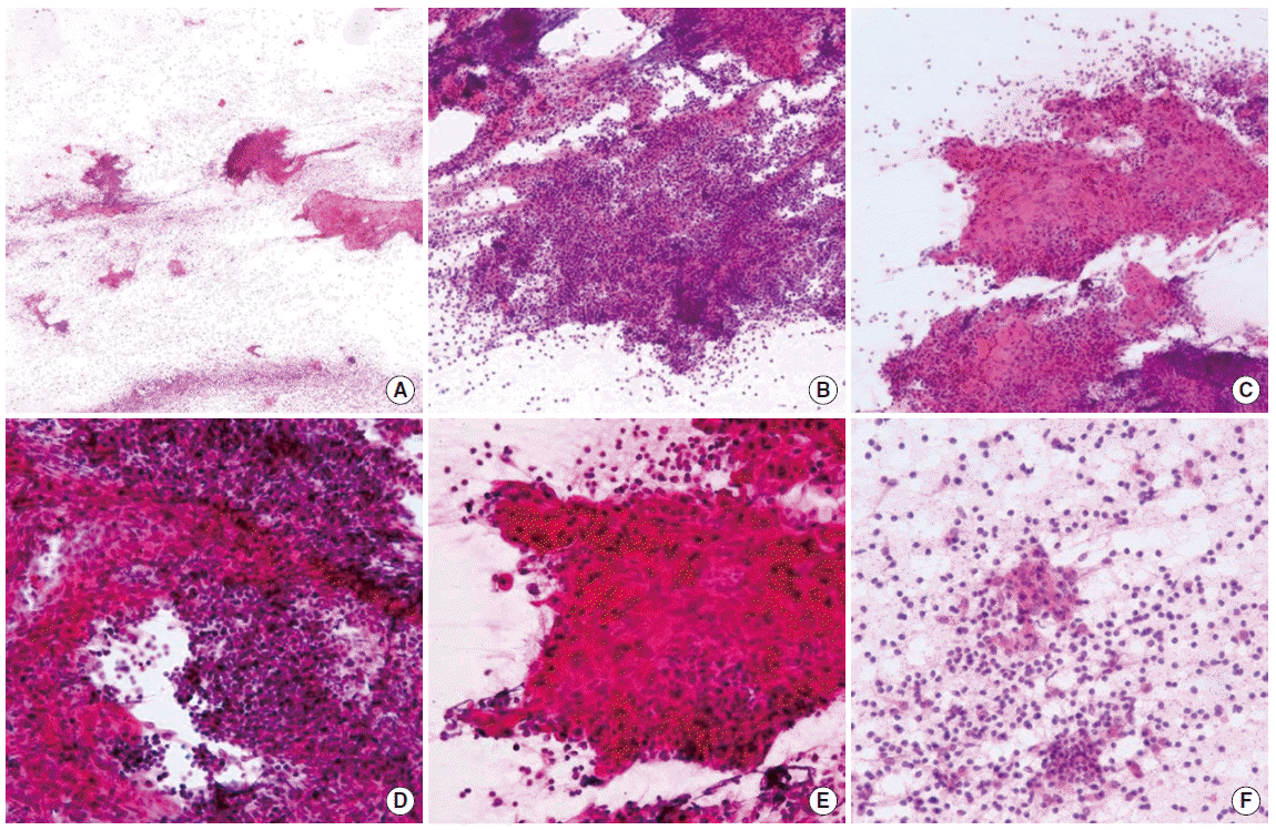
Fig. 2.
Fine needle aspiration cytology of the lymphoepithelial cyst of the thyroid gland. (A) Syncytial tissue fragments are present in the lymphocytic background. (H&E, × 12.5). (B) Cellular fragments show papillary-like configuration with fibrovascular stroma (H&E, × 40). (C) Area suggesting squamous metaplasia is identified in the tissue fragments (H&E, × 100). (D) Squamous components (left side) and clusters of lymphoid cells (center and superior side) are closely blended (H&E, × 200). (E) Some areas of squamous cells show unorganized arrangement and mild cellular atypia (H&E, × 400). (F) Several small solid cell nests and detached cells are intermingled with lymphocytes (H&E, × 400).