투석 중에 발생한 신경병증 양상의 통증에서 초음파 유도하 신경 수력분리술을 이용한 치료: 증례보고
Ultrasound-Guided Nerve Hydrodissection for Neuropathic-Like Pain Arising from Hemodialysis: A Case Report
Article information
Trans Abstract
Peripheral neuropathy is very common in patients with chronic renal failure. The pain arising from hemodialysis can be caused by vascular problems (such as vascular stenosis and steal syndrome) and neuropathy. Hemodialysis patients who need to be dialyzed three times a week should not be told to endure worsening pain every time they are dialyzed. We report that the pain arising from hemodialysis in the lower arm was a concern due to the nerve entrapment in the axillary area, and it was successfully controlled with ultrasound-guided nerve hydrodissection.
서 론
Marzouq 등[1]은 투석 환자들 중 66.3%가 만성 통증을 가지고 있고, 약물치료를 받아도 26.6%는 전혀 효과가 없었다고 보고했다. 특히 말초신경병증은 만성신부전 환자에서 매우 흔하다. Gondhali 등[2]은 말초신경병증이 만성신부전 환자 중에서도 투석 전 환자와 비교하여 투석 환자에서 더 흔하고, 만성신부전의 병기(stage)와 유병기간(duration of disease)이 증가함에 따라 빈도와 중증도가 증가한다고 보고한 바 있다.
투석 중 발생하는 통증은 투석혈관의 협착, 혈관 스틸(steal syndrome) 등과 같이 혈관의 문제와 신경병증이 원인일 수 있다. Harris 등[3]은 만성신부전 환자에서 신경병증을 3가지로 분류하여 기술하였다. 첫 번째, 당뇨병성 신경병증(diabetic polyneuropathy), 요독성 다발성 신경병증 같은 전신적 질환과 관련한 신경병증(systemic disease neuropathy)과 두 번째, 손목터널증후군(carpal tunnel syndrome), 팔꿈치터널증후군(cubital tunnel syndrome), 척골관증후군(Guyon’s tunnel syndrome)처럼 압박에 의한 경우와 구획증후군(compartment syndrome), 또는 신경포착(nerve entrapment) 에 의한 단일신경병증(mononeuropathy), 그리고 세 번째, 허혈성 일측 사지 신경병증(ischemic monomelic neuropathy) 등이다.
본 증례는 투석 중 발생한 신경병증 양상의 통증으로 투석혈관 확장으로 인한 신경포착이 원인으로 의심되어 5% dextrose water (5%DW) 수액으로 신경 수력분리술(nerve hydrodissection)을 시행하여 효과적으로 치료되어서 보고하고자 한다.
증 례
70세 남자는 고혈압이 있었고 당뇨는 없었다. 만성신부전 환자로 투석을 위한 접근로(hemodialysis access) 확보를 위해 우측 요골 동맥(radial artery)과 요골측 피부정맥(cephalic vein)을 연결하는 동정맥루 형성술(arterio-venous fistula formation)을 받았다. 수술 6주 후에도 요골측 피부정맥이 충분히 성숙되지 않아 우측 척골측 피부정맥(basilic vein)을 전위시켜 상완동맥(brachial artery)과 연 결하는 형성술(basilic vein transposition)을 받았다. 수술 4주 후에 혈관에 문제가 없어 투석접근로(hemodialysis access)로 사용하였는데, 투석 시작 후 2시간 정도 지나면 우측 아래팔의 척골측 부위에 찌릿한 양상의 통증(paresthesia)이 발생하였고 투석이 진행되는 동안 계속 악화되다가(Visual Analog Scale [VAS] score=58) 투석이 끝나면 그 통증은 사라졌다고 하였다. 내원 당시 근력약화 소견은 없었고, 감각둔화(numbness)와 바늘통각검사(pin prick test)에서 양성 소견을 보였으며 그 외 이상소견은 없었다. 통증이 수술 후 4주가 지나서 발생했고 투석 시에만 발생하는 통증이라 수술과정에서 발생한 신경 손상과 허혈성 일측 사지 신경병증은 가능성이 낮다고 판단하여 근전도검사는 따로 시행하지 않았다.
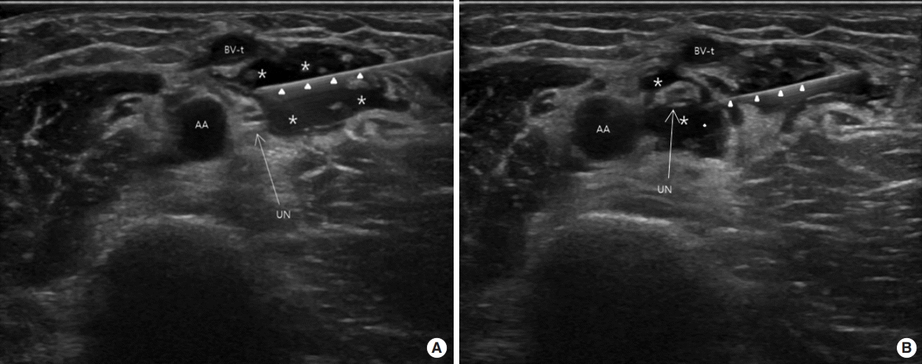
통증 부위의 지배 신경을 초음파(LOGIQ P9; GE Healthcare, Chicago, IL, USA; linear probe, 5–15 MHz)로 원위부에서 쇄골하 부위(infraclavicular area)까지 추적 관찰했을 때 통증의 가장 근위부인 겨드랑이 위치에서 동맥과 정맥 사이에 있는 척골신경의 다발(nerve fascicle)이 커져 있는 것이 확인되어 반대쪽 상지의 신경과 비교하였다(Fig. 1). 투석 중 혈관 확장으로 인한 신경자극이 통증의 원인일 것으로 추정되어 신경 수력분리술을 시행하였다. 바늘 진입 시 혈관 손상을 피하기 위해 초음파 탐색자(probe)를 원위 부위(distal) 방향으로 이동하여 척골신경이 노출되는 부위에서 실시간 초음파(LOGIQ P9; GE Healthcare; linear probe, 5–15 MHz) 유도하에 26G, 5.08 cm 바늘을 긴 축(in-plane)으로 진입시켰다. 바늘 끝 경사면을 계속 확인하면서 바늘을 진입시켰고, 바늘 끝이 신경외막(epineurium) 근처에 닿았을 때 주사액 5%DW 12 mL (20%DW 3 mL+1% lidocaine 4 mL+normal saline 5 mL)를 척골신경 위쪽과 아래쪽에 주입하면서 신경과 주위 조직을 박리하였다(Fig. 2). 바늘 진입 시 이상감각(paresthesia)은 발생하지 않았으며, 주사 완료 후 신경은 무에코성 고리(anechoic ring; a black halo)를 가지고 있는 것처럼 보였다[4,5] (Fig. 3). 통증의 평가는 VAS score (0–100)로 하였으며, 내원 당시 통증은 없는 상태라 환자 기억에 의존하여 투석 당시 통증을 평가하였다. 시술 후 1주일째에 투석 시 발생했던 통증이 사라졌고(VAS score=0), 주사로 인한 합병증(감염, 혈종, 신경 손상 등)도 나타나지 않았으며, 6개월이 지난 지금까지 통증이 재발되지 않았다. 본 증례를 보고하기 위해 환자에게 의무기록을 포함한 의학정보의 연구자료 이용 동의를 받았다.

Ulnar nerve (UN) in the right side (A) is larger than left side (B) and nerve fascicles are swelling. AA, axillary artery; AV, axillary vein; BV-t, transposed basilic vein.

(A, B) In order to avoid vessel damage during needle injection, (B) the probe was moved in the distal direction to expose the ulnar nerve (UN). The axillary vein (AV) seen in (A) is not visible. The tip of the needle is seen near the UN (arrowhead). Paresthesia was not provoked. Asterisks (*) indicate anechoic injectant; white arrowheads indicate needle. AA, axillary artery; BV-t, transposed basilic vein.

(A) First, injectant was injected into the upper part of the ulnar nerve (UN), and (B) then into the lower part of the UN. The UN is completely separated from the surrounding tissue and floating. An anechoic ring (a black halo) is observed. Asterisks (*) indicate anechoic injectant; white arrowheads indicate needle. AA, axillary artery; BV-t, transposed basilic vein.
고 찰
평소에 통증이 없다가 투석 시에만 통증이 발생하는 경우, 투석 시작하자마자 또는 1시간, 2시간, 3시간 후에도 발생 가능하며, 통증 부위는 어깨, 위팔, 아래팔, 손가락 등 투석혈관이 있는 상지 쪽에 주로 발생한다. 매주 3회 투석해야만 하는 투석 환자들에게 투 석할 때마다 악화되는 통증을 마냥 참으라고만 해서는 안 된다. 본 연구과정에서도 도저히 투석을 하지 못하고 3시간 이내 투석을 중단하는 사례들을 볼 수 있었으며, 조절되지 않는 통증으로 새로 혈관수술을 받는 경우도 있었다. 물론 평소에는 통증이 없기 때문에 통증을 정확히 표현하지 못하는 경우가 많아 원인을 감별하기가 쉽지 않다. 반대편 상지에도 드물게 통증을 호소하는데, 이런 경우 투석혈관과 무관하게 환자가 이전에 가지고 있던 근골격계 및 신경계 질환에 의한 통증이 투석시간 동안 고정된 자세에 의해 악화되는 것으로 보인다.
포착된 신경의 식별은 반대쪽과 비교해서 부어 있는 경우 또는 신경 내 여러 신경다발(nerve fascicles)들 중 커져 있는 다발이 있는 경우와 직접 의심되는 신경을 촉진했을 때 환자가 경험했던 통증이 재현되는 경우에 가능하다[4]. 하지만 투석혈관이 확장되면서 정상 해부학적 구조가 변형되어 신경 위치를 찾기 어려울 수 있다. 특히 투석을 안 할 때는 신경포착의 증상도 없기 때문에 촉진 및 초음파검사로도 확인이 어려울 수 있다. 따라서 신경포착에 의한 통증이 의심된다면 투석 시작 전과 투석 중 통증 발생 시 혈관이 어떻게 변하는지 투석실에서 실시간 초음파검사를 시행하는 것이 감별 진단에 도움이 되리라 생각한다.
신경포착으로 인한 통증은 포착된 신경의 분포영역에 이상감각(paresthesia), 통각과민(hyperalgesia), 무감각(numbness), 저림증상(tingling sensation) 등 신경병증 양상을 보인다. 그래서 다른 원인의 신경병증과 감별이 필요한데, 특히 허혈성 일측 사지 신경병증과 임상 양상이 비슷해서 적절한 치료시기를 놓치게 되면 돌이킬 수 없는 신경학적 결손을 초래하기 때문에 주의를 필요로 한다. 허혈성 일측 사지 신경병증은 투석혈관 형성 환자 1% 이하에서 발생하는 것으로 추정되며, 주로 투석혈관 형성 직후에 발생하고, 말초신경병증을 동반한 당뇨병 환자, 여성 환자, 그리고 상완동맥(brachial artery)에서 투석혈관을 연결했을 때 많이 발생한다[6].
신경포착에 의한 신경병증 치료에서 신경 수력분리술의 효과에 대해 연구가 많이 진행되고 있다[7]. 신경포착은 약한 압박(mild compression)에 의해서도 신경외막(epineurium)에 분포하는 신경벽 신경(nervi nervorum), 신경속 혈관(vasa nervorum)에 영향을 줘 통증을 발생할 수 있으며, 신경 수력분리술은 신경 주위에 압박될 수 있는 구조물로부터 신경을 유리시켜 신경을 안정시키는 치료법이다[8]. 분리술에 사용되는 약물의 선택에 대해서는 더 연구가 필요하겠으나 신경병증 통증(neuropathic pain)에서 5%DW의 효과에 대해 많이 연구되고 있다. Chao 등[8]은 손목터널증후군(carpal tunnel syndrome)에서 5%DW를 이용한 신경 수력분리술이 효과가 있음을 보고하였고, Stoddard 등[9]은 주관절터널증후군(cubital tunnel syndrome)에서 5%DW만으로 신경 수력분리술을 시행하여 효과적으로 치료한 증례를 보고하였다. 또한 Maniquis-Smigel 등[10]은 하지 방사통을 동반한 척추질환 환자에서도 경막외공간(epidural space)에 5%DW 주입이 효과 있음을 보고한 바 있다. 5%DW를 이용한 신경 수력분리술은 구조적 압박을 풀어주는 효과뿐만 아니라 5%DW 자체가 통증을 완화해주는 효과가 있으며, 과민반응과 과량 사용에 따른 부작용이 적어 본 증례에서는 5%DW 수액과 국소 마취제(lidocaine) 혼합액을 사용하였다.
신경 수력분리술은 신경과 혈관 및 주위 조직 사이를 분리시키는 주사요법으로 시술과 관련된 여러 합병증들이 있으며, 그 중 심각한 합병증으로는 감염(infection), 신경 내 주사(intraneural injection) 그리고 혈종(hematoma), 가성 동맥류(pseudoaneurysm) 같은 혈관의 손상으로 인한 것 등이 발생할 수 있다. 특히 투석 환자들은 투석혈관의 확장으로 해부학적 구조가 변형된 경우가 많아 반드시 초음파 유도하에 주사 치료를 시행하여야 하며, 안전하게 주사 치료하기 위해서는 숙련과정이 필요하다.
결론적으로, 투석과 관련하여 발생하는 통증은 원인을 정확히 진단하는 것이 무엇보다도 중요하다. 특히 혈관 협착, 혈관 스틸(steal syndrome), 허혈성 일측 사지 신경병증(ischemic monomelic neuropathy), 가성 동맥류 형성 및 혈종에 의한 신경압박 증상 같은 빠른 수술적 처치가 필요한 경우 등이 감별되어야 한다. 본 증례에서는 투석 중 발생한 아래팔 부위의 통증에 대해 초음파 유도하 신경 수력분리술이 만족할 만한 효과가 있었다. 하지만 정확한 진단과 치료를 위해 향후 통증 부위와 신경포착 부위에 대한 관련 연구가 더 필요하리라 생각된다.