경정맥공에 발생한 판상 수막종의 증례보고 및 감별진단의 영상소견 비교
A Case of En Plaque Meningioma of Jugular Foramen with Image Findings of Differential Diagnosis
Article information
Trans Abstract
Primary meningioma of jugular foramen is extremely rare, while paraganglioma or nerve sheath tumor are relatively common in jugular foramen. We reported a case of primary meningioma of jugular foramen. A 79-year-old female who had left tinnitus and hearing loss for three month came to the department of otorhinolaryngology. Temporal bone computed tomography scan showed sclerotic change and slightly irregular margins of left jugular foramen with relatively preservation of bony architecture. Temporal bone magnetic resonance image showed well defined homogeneous enhancing mass in left jugular foramen with extension to carotid space on gadolinium enhanced T1 weighted image. Prominent dural tail was also noted. On T2 weighted image, this mass showed intermediated signal intensity with no vascular signal voids. Meningioma was confirmed by pathology. In this article, we describe a case of primary en plaque meningioma of jugular foramen and review image findings of differential diagnosis.
서 론
경정맥공에서 발생하는 일차성 종양들 중 가장 흔한 것은 경정맥공 사구종(paraganglioma)으로 그 빈도는 약 90%이다. 그 다음으로는 신경피막 종양을 들 수 있으며 그 중에서도 신경초종(schwannoma)이 이에 해당한다. 반면 경정맥공에서 일차적으로 발생한 수막종(meningioma)은 매우 드물고 전체 후두와(posterior fossa)에서 발생하는 수막종 중 0.7%–9.3%를 차지한다. 경정맥공은 경정맥, 9번, 10번, 11번 뇌신경을 포함하는 복잡한 해부학적 구조물로서 일차성 종양이 발생하게 되면 이와 관련된 뇌신경 이상증상을 보인다[1]. 저자들은 이명과 청력소실, 두통을 주소로 내원하였던 79세 여자 환자에서 왼쪽 경정맥공(jugular foramen)에 발생하여 경동맥공간(carotid space)까지 확장되어 있는 일차성 판상(en plaque) 수막종의 증례를 경험하였으며 문헌고찰과 함께 이에 대한 특징적인 영상소견 및 경정맥공에 생긴 사구종과 신경초종과의 영상의학적 차이점을 기술하고자 한다.
증 례
79세 여자 환자이며 3개월 전부터 발생한 왼쪽 이명과 청력소실, 두통을 주소로 순천향대학교 천안병원 이비인후과에 내원하였다. 과거력상 고혈압 외에는 특이사항은 없었다. 측두골 전산화단층촬영(computed tomography)과 측두골 자기공명영상(magnetic resonance imaging)을 시행하였고 측두골 전산화단층촬영에서 왼쪽 경정맥공의 경화성 골변화 및 불균일한 테두리를 보였으며 골구조는 비교적 유지되어 있는 반면 골미란의 소견은 없었다. 또한 왼쪽 중이강과 유양동 내에 미만성 음영증가소견이 보였으며 일부 약간의 경화성 골변화 외에 공기음영이 대부분 소실되어 있었다(Fig. 1A). 측두골 자기공명영상에서는 왼쪽 소뇌교각부에 넓은 기저부를 두고 있는 균질한 조영증강의 종괴가 가돌리늄(gadolinium) 조영증강 T1 강조영상에서 보였으며 경질막 꼬리 징후(dural tail sign)를 동반하고 있고 판상형태를 보였다(Fig. 1B). 이 종괴는 T1와 T2 강조영상에서는 중등도의 신호를 나타내고 왼쪽 경정맥공을 중심으로 왼쪽 경동맥공간까지 미만성으로 확장되어 있는 양상이었다(Fig. 1C). 또한 추체골(petrous bone)과 경사대(clivus)의 왼쪽 부위에는 T2 강조영상에서 저신호를 보이며 가돌리늄 조영증강 T1 강조영상에서 비균질한 조영증강소견을 보였다(Fig. 1D). T2 강조영상에서 종괴 내에 혈관 신호소실(vascular signal void)은 보이지 않았다. 이 종괴에 대하여 경유양돌기 접근법(transmastoid approach)을 통하여 조직검사를 시행하였고 수막종으로 진단되었다.

79-year-old female with en plaque meningioma of jugular foramen. (A) Bone window image from temporal computed tomography scan shows sclerotic change and slightly irregular margins of left jugular foramen (arrow) with relatively preservation of bony architecture, resulting in characteristic permeative sclerotic appearance. (B, C) Gadolinium enhanced T1 weighted images show well defined homogeneous enhancing mass in left jugular foramen with en plaque involvement of posterior fossa and carotid space. Note prominent dural tail sign (arrow). (D) Gadolinium enhanced T1 weighted image shows heterogeneous enhancement of left petrous bone and clivus, which means tumor spread. A, anterior; P, posterior.
고 찰
경정맥공에서 발생하는 일차성 종양들 중 가장 흔한 것은 경정맥공 사구종으로 그 빈도는 약 90%이다. 그 다음으로는 신경피막종양을 들 수 있으며 그 중에서도 신경초종이 이에 해당한다. 경정맥공에서 일차적으로 발생한 수막종은 매우 드물고 전체 후두와 수막종의 0.7%–9.3%를 차지한다. 경정맥공은 경정맥, 9번, 10번, 11번 뇌신경을 포함하는 복잡한 해부학적 구조물로서 일차성 종양이 발생하게 되면 이와 관련된 뇌신경 이상증상을 보인다[1]. 미각의 소실이나 성대마비나 삼킴곤란, 목빗근(sternocleidomastoid muscle)과 승모근(trapezius muscle)의 쇠약이 나타날 수 있으며 청각경로(auditory pathway)를 침범하거나 뇌줄기, 소뇌를 압박할 경우 이명, 청력소실, 안진, 보행장애, 어지러움 등의 증상이 나타난다[2].
경정맥공에서 일차적으로 발생하는 수막종(일차성 수막종)은 이차적으로 발생하는 수막종(이차성 수막종)과는 다른 양상을 나타낸다. 이차성 수막종의 경우 골침윤은 제한적이며 주로 연부조직 종괴를 형성하게 되는 반면 경정맥공의 일차성 수막종은 국소적인 골침윤 경향이 더 심하다. 또한 이러한 침윤은 모든 방향으로 나타나게 되고 이러한 패턴의 침윤을 원심성(centrifugal) 침윤이라고 한다. 바깥쪽으로는 측두골의 중이강, 안쪽으로는 경정맥공 결절(jugular tubercle), 혀밑신경관(hypoglossal canal), 뒤통수뼈관절융기(occipital condyle) 등을 침윤할 수 있으며 위쪽으로는 뇌경질막(intracranial dural reflection)을 따라서 두개골 내까지 침윤할 수 있다. 특히 이러한 양상의 경질막을 따라 보이는 침윤에 대하여 판상(en plaque)이라고 지칭하며 원심성 침윤과 함께 이는 일차성으로 발생하는 경정맥공 수막종의 특징이다. 반면 감별질환인 경정맥공 사구종의 경우 경정맥공으로부터 위가쪽 방향으로 침윤하며 중이강의 고실하부를 주로 포함한다[3]. 감별질환 중의 하나인 신경초종은 대부분 8번 뇌신경의 위안뜰가지(superior vestibular branch)가 위치하는 소뇌교각부에 흔히 발생하지만 드물게 경정맥공 내의 9번, 10번 뇌신경으로부터 발생할 수 있다[4]. 신경초종은 신경의 주행을 따라 발생하기 때문에 주로 위안쪽 방향으로 침윤하게 된다[3].
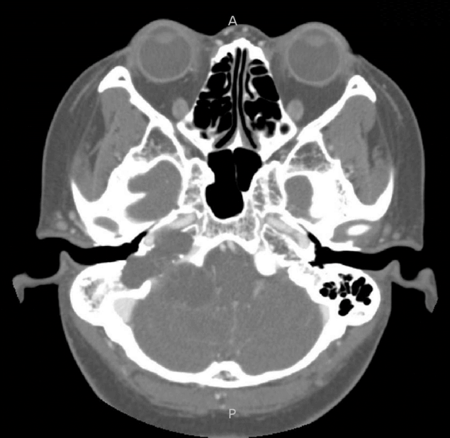
경정맥공 사구종이나 신경초종을 포함한 다른 감별질환들을 진단하는 데 있어서 전산화단층촬영과 자기공명영상 모두 중요한 역할을 한다. 비록 자기공명영상이 주변조직과의 관계가 명확하여 정확한 종양의 위치 및 범위를 알 수 있게 해준다는 점에서 더 선호되지만 전산화단층촬영 또한 종양과 골조직과의 관계를 더 명확하게 알려주고 수술자에게 해부학적 기준점을 알려줄 수 있기 때문에 매우 중요하다고 할 수 있다[5]. 전산화단층촬영에서 경정맥공 수막종은 경정맥공에 중심을 두고 중이강으로 퍼져나가는 양상의 종괴로 특징적으로 골조직의 침습경화 변화(mixed permeative and sclerotic bone change)를 보인다[6]. 종괴의 직접적인 침윤으로 인해 현저한 골과다(hyperostosis) 또는 골경화를 보이며 정상 골피질의 소실과 함께 불균일한 골테두리를 보이지만 상대적으로 골구조는 유지되어 있고 골미란 등의 소견은 보이지 않는다는 점이 특징이다[3]. 반면 경정맥 사구종은 골조직의 침습파괴 변화(permeative and destructive bone change)를 보이게 된다는 점에서 감별할 수 있다. 즉 골구조가 유지되어 있지 않으며 골파괴 및 골미란 소견을 보인다(Fig. 2). 신경초종의 경우 종괴의 크기가 점차적으로 증가함에 따라서 압박미란(pressure erosion)에 의해 경정맥공의 크기가 커지지만 경정맥공의 골피질이 잘 유지되어 있다[1]. 따라서 경정맥공의 용해성 골파괴보다는 조개모양의(scalloped) 경화성 골 확장을 보이게 되는 점이 특징이다[4] (Fig. 3).

65-year-old male with paraganglioma of jugular foramen. Bone window image from temporal computed tomography scan shows extensive bone destruction and erosion of margins of right jugular foramen and temporal bone. P, posterior.
35-year-old female with schwannoma of jugular foramen. Bone window image from temporal computed tomography scan shows sclerotic expansion of right jugular foramen with scalloped appearance and preservation of cortex. A, anterior; P, posterior.
자기공명영상에서 경정맥공 수막종은 원심성의 두개저 침윤을 보이며 침윤된 두개저는 가돌리늄 T1 강조영상에서 다양한 조영증강소견을 보이게 되고 특징적으로 골구조는 잘 유지되어 있다. 또한 두개 내 조영증강되는 경질막 꼬리 징후를 볼 수 있으며 혈관 신호소실이 보이지 않는다는 점이 특징적이다. 반면 경정맥 사구종의 경우 혈관 신호소실을 보이는 점이 특징적이라고 할 수 있다[6]. 신경초종의 경우 T1 강조영상에서 저신호, T2 강조영상에서 고신호 강도를 보이며 혈관 신호소실이나 경질막 꼬리징후는 보이지 않는다[4].
하지만 비전형적인 영상소견을 보이는 경정맥공 수막종의 경우 경정맥공 사구종이나 신경초종과의 감별이 어려울 수 있다. Chen 등[2]이 보고한 혈관종성(angiomatous type) 경정맥공 수막종의 경우 저명한 골파괴소견을 보였으며 자기공명영상에서 낭성변화로 인한 소금후추가루 양상(salt and pepper pattern)을 보였다. 경정맥공 사구종과 신경초종의 경우에도 비전형적인 영상소견을 보이는 경우 두 질환의 감별이 어려울 수 있다. Lee 등[4]이 보고한 증례의 경우 왼쪽 경정맥공에 생긴 골용해성의 종괴로 사구종을 모방하였지만 신경초종으로 진단되었다. 순천향대학교 천안병원에서의 또 다른 증례에서도 양측 경동맥공간과 경정맥공에 보이던 종양으로 현저한 골파괴소견을 동반하고 있어 사구종을 의심하였지만 신경초종으로 확인되었던 바 있다(Fig. 4).

56-year-old female with atypical schwannoma mimicking paraganglioma. (A) Bone window image from temporal computed tomography scan shows extensive destructive bone change of both jugular foramen. (B) Gadolinium enhanced T1 weighted image shows homogeneous enhancing mass in both jugular foramen with involvement of posterior fossa and carotid space. Histologic diagnosis revealed schwannoma. A, anterior; P, posterior.
본 증례에서는 전산화단층촬영에서 골미란이나 골파괴가 동반되어 있지 않은 종양으로 왼쪽 경정맥공의 경화성 골변화가 있으며 가돌리늄 조영증강 T1 강조영상에서는 왼쪽 경정맥공을 중심으로 경동맥공간까지 미만성으로 확장되어 있는 균질한 조영증강의 종괴가 보였다. 종괴는 T2, T1 강조영상에서 중등도의 신호 강도를 보이며 추체골과 경사대는 골침윤으로 인해 T2강조영상에서 저신호강도를 보이고 있었고 경정맥공과 뇌기저부뿐만 아니라 경동맥공간까지 확장되어있는 소견을 보였다. 가돌리늄 조영증강 T1 강조영상에서 두개 내 조영증강되는 경질막 꼬리징후가 명확하였고 T2 강조영상에서 혈관신호 소실이 보이지 않았다. 따라서 이러한 특징으로 보았을 때 이전 문헌들에서 언급되었던 경정맥공 수막종의 영상 소견과 일치하는 것으로 생각하였다. 경동맥공간까지 확장되어 있다는 점이 자칫 경정맥공 사구종이나 신경초종의 가능성을 생각할 수 있게 하지만 일차성 경정맥공 수막종의 증례들을 보고하였던 이전의 문헌에서도 확인할 수 있듯이 수막종에서도 흔한 소견이었다[3]. 경동맥공간 내에 국한된 종양은 수막종의 가능성이 매우 적으나 경정맥공과 경동맥공간을 포함하여 확장되어 있는 종양은 수막종의 가능성을 배제할 수 없다.
경정맥공에서 발생하는 일차성 수막종은 경정맥공 사구종이나 신경초종에 비해 그 빈도가 드물지만 전산화단층촬영과 자기공명영상의 특징적인 영상소견으로 감별이 가능하다. 즉 두개저를 침범하는 원심성의 종괴로서 전산화단층촬영에서 골구조의 파괴가 동반되어 있지 않은 침습경화 변화를 보이고 자기공명영상에서 경질막 꼬리징후를 보이며 혈관 신호소실은 보이지 않는다. 따라서 이러한 영상소견을 보일 때 경정맥공 수막종을 진단하는 데 도움이 될 수 있다.