Recurred Segmental Schwannomatosis Without Neurofibromatosis Type 2
Article information
Abstract
Schwannomas are the most common type of benign peripheral nerve sheath tumors. They typically present as a solitary lesion, but multiple schwannomas rarely occur in patients with neurofibromatosis type 2 (NF2), or patients without the other hallmarks of NF2. The latter is termed schwannomatosis. They most commonly occur in the head and neck involving the brachial plexus and spinal nerves. Although rarely found in the extremities, when these masses occur peripherally, they most commonly affect the sciatic, ulnar, and tibial nerve. It is reported that 2.4% to 5% of all patients undergoing schwannoma excision present as schwannomatosis. One-third of patients with schwannomatosis show tumors limited to a single extremity or segment of the spine and it is referred to as segmental schwannomatosis. We report a case of recurred segmental schwannomatosis of the posterior tibial nerve without features of NF2 after schwannoma excision.
INTRODUCTION
Schwannomas are the most common type of benign peripheral nerve sheath tumors originating Schwann cells [1,2]. Several names have been used to describe the tumor, including peripheral glioma, perineurial fibroblastoma, schwannoma, neurinoma, and neurilemoma. Today the two most frequently used terms in the literature are neurilemoma and schwannoma [3]. Although more commonly appearing in isolation, 5% of these tumors grow in a plexiform or multinodular pattern [2]. While multiple schwannomas are typically associated with neurofibromatosis type 2 (NF2), they may occur without pathognomonic bilateral vestibular nerve involvement and classic ophthalmological and dermatological stigmata, in which case the condition is termed schwannomatosis. Schwannomatosis is recognized as the third major form of neurofibromatosis with a specific set of diagnostic criteria [4,5]. No estimate of the prevalence of schwannomatosis has been reported, but annual incidence was estimated to be approximately 1/1,700,000 in a Finnish population-based study [5]. One-third of patients with schwannomatosis show tumors limited to a single extremity or segment of the spine and it is referred to as segmental schwannomatosis [4].
We report the case of a 30-year-old woman with recurred segmental schwannomatosis affecting the posterior tibial nerve without the other hallmarks of NF2 after schwannoma resection.
CASE REPORT
A 30-year-old woman presented with a 2-year history of palpable painful nodules on the medial and lateral aspect of her left ankle and foot. She had no other signs, symptoms, systemic disease, or family history of neurofibromatosis. She stated the nodule had been solitary, small, and asymptomatic in the beginning but had gradually increased size and number. The patient denied any significant trauma to the affected foot. She underwent previous surgery for excision of benign schwannoma confirmed histologically from the medial aspect of the foot 10 years ago.
Physical examination revealed three palpable nodules with tenderness in the medial and lateral aspect of the left foot and ankle, but without swelling. The nodules were firm, palpable, and painful on direct palpation. A scar, corresponding to the site of the previous foot mass resection, was also noted. Otherwise, she had well-preserved foot and ankle alignment. Neurological examination showed no muscle atrophy, and there was a full range of movement. Light touch and vibratory sensations were normal. There was no Tinel’s sign. Routine laboratory results showed no remarkable findings.
Plain radiographs showed no osseous deformity or lesion (Fig. 1). Magnetic resonance image (MRI) was performed to characterize the nodules detected during physical examination. MRI demonstrated multiple, well-defined, round, or fusiform-shaped nodules along the course of the posterior tibial nerve and their branches, with segmental distribution and characteristics suggestive of peripheral nerve sheath tumors. Separate nodules ranged from 0.5 to 2 cm in maximum dimension. Lesions were heterogeneously hyperintense on fat-suppressed T2-weighted images (Fig. 2A) and isointense to muscle on T1-weighted images. Heterogeneous intense enhancement was identified after gadolinium administration (Fig. 2B, C). Based on the clinical and imaging findings, it was highly likely that nodules represented a peripheral nerve sheath tumors arising from the posterior tibial nerve.

Radiograph of the left ankle shows no osseous deformity or lesion.
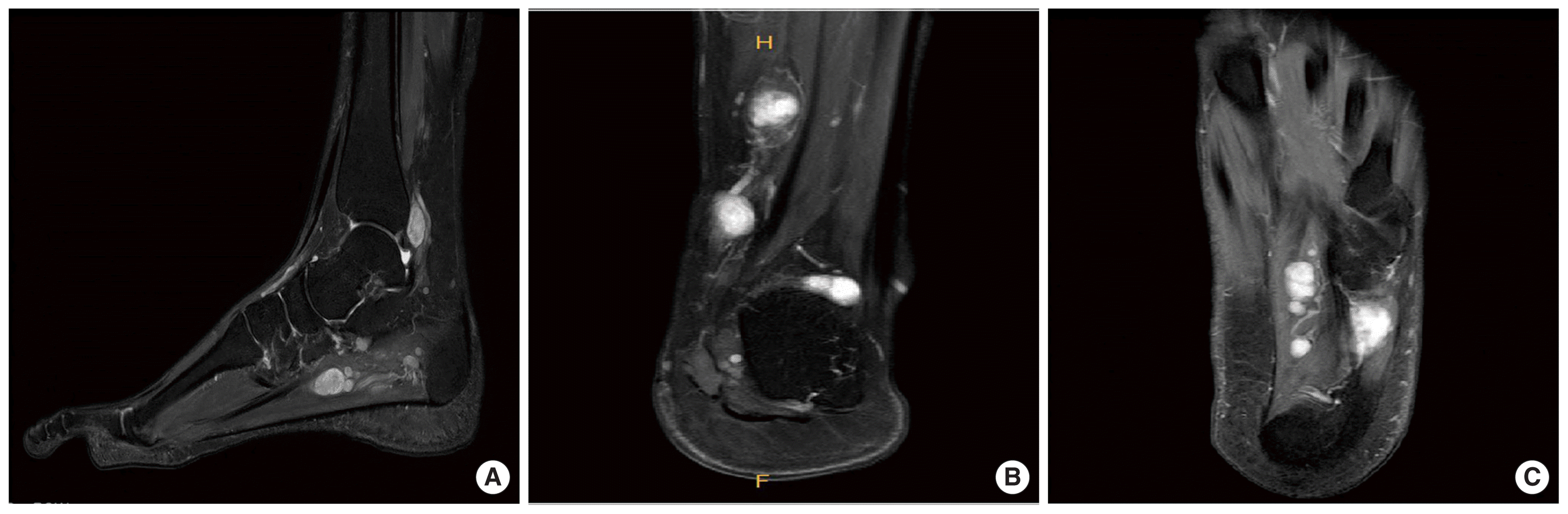
Magnetic resonance imaging demonstrates multiple round and fusiform-shaped nodules along the course of the posterior tibial nerve and their branches, ranging from 0.5 to 2 cm in maximum dimension. (A) On sagittal fat-suppressed T2-weighted image, these nodules show heterogeneously hyperintense signal intensity. (B) Coronal and (C) axial gadolinium enhanced T1-weighted images show hetereogeneous intense enhancement of nodules.
The patient underwent surgery under general anesthesia. An incision was made over the lesions with care to avoid the marked vascular structures and the nodules were identified and carefully excised, taking care to leave it intact while avoiding damage to the posterior tibial nerve.
Grossly, the resected specimen measured 2.0×1.5×0.5 cm of medial aspect, 3.5×1.5×0.5 cm in aggregates of lateral aspect. Histologically, the specimen was an encapsulated lesion with proliferating spindle cells in a palisade arrangement (Fig. 3). An immunostain for S-100 protein showed the strong, uniform reactivity characteristic of schwannoma.
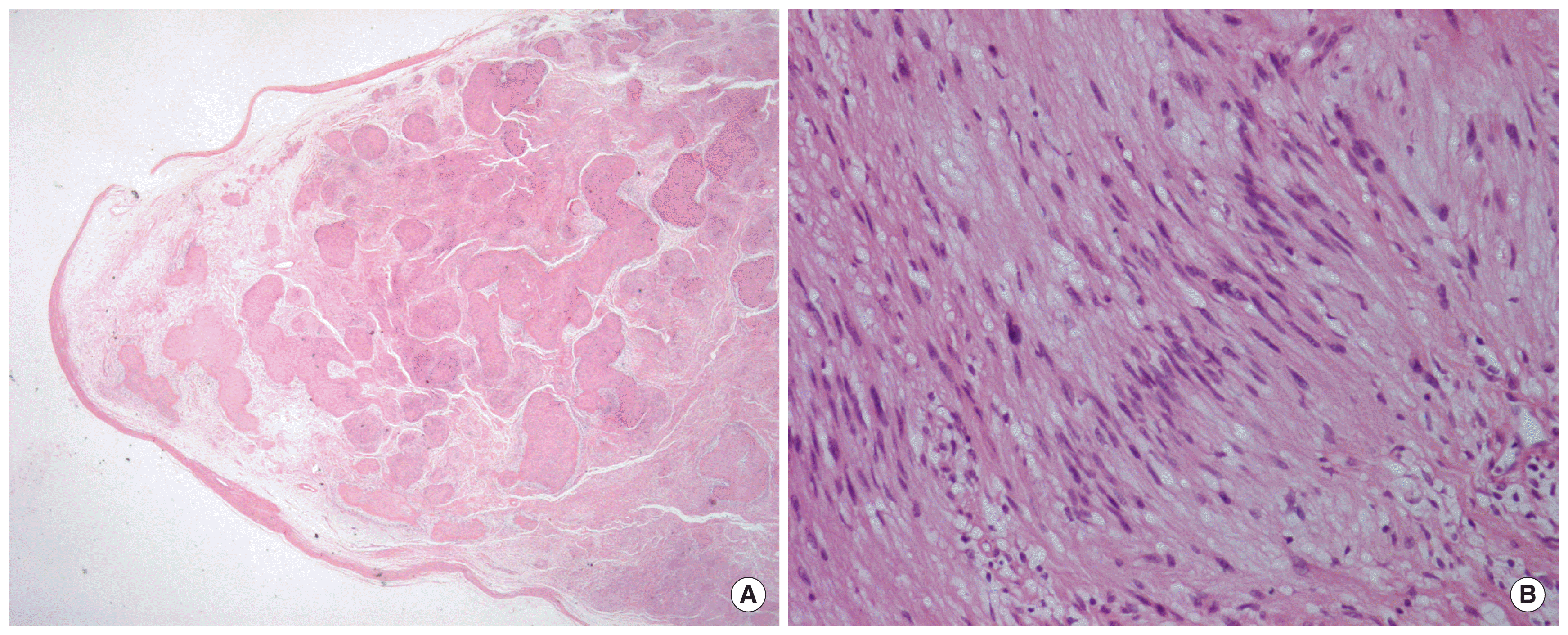
(A) The specimen shows encapsulated lesion with mixed cellular (Antoni A) and loose hypocellular (Antoni B) area (H&E, × 4). (B) The cellular Antoni A area shows characteristic Verocay body of nuclear palisading and cell-free stroma (H&E, × 200).
DISCUSSION
Multiple schwannomas are slow growing encapsulated tumors of the nerve sheaths. Although they can be found anywhere in the body, they have a predilection for the head, neck, and flexor surfaces of extremities because their preferred localization is nerve roots. Although rarely found in the extremities, when these masses occur peripherally, they most commonly affect the sciatic, ulnar, and tibial nerve. Clinically, they usually represent as slow growing solitary masses, but approximately 5% of these tumors grow in a plexiform or multinodular pattern [2,4]. Multiple schwannomas should raise suspicion for a diagnosis of neurofibromatosis, but recently it has been recognized that some patients with multiple schwannomas lack vestibular tumors. This condition constitutes the third major form of neurofibromatosis termed schwannomatosis which may be as common as NF2 [6]. As it is of low incidence, schwannomatosis contributes to the difficulty in obtaining an accurate diagnosis. Because schwannomatosis shows considerable differences in anatomic distribution of lesions and clinical presentation, medical management, and patient outcomes, despite the overlap between schwannomatosis and NF2 in terms of presentation and phenotype, it is recognized as a separate clinical entity.
Schwannomatosis is a rare disorder of unknown prevalence. The reported incidence of schwannomatosis ranges from 1/40,000 to 1/1,700,000, suggesting an incidence similar to that of NF2 [7,8]. It is also reported that 2.4% to 5% of all patients undergoing schwannoma excision present as schwannomatosis [4]. There is no sex predilection and the most common age group is between 30 and 60 years which is similar to that of schwannoma and older than that of NF2 [2].
MRI is the most useful modality in work-up and diagnosis of schwannomatosis. According to several studies, the MRI finding of schwannomatosis is characterized by multiple discrete, well-defined, rounded, or oval lesions distributed along the courses of peripheral nerves in the extremities and in the paraspinous nerve roots. Schwannomatosis shows signal characteristics that resemble isolated schwannomas; they are typically low to intermediate signal intensity on unenhanced T1-weighted images, high signal intensity on proton density, T2-weighted, and STIR images and heterogeneous enhancement after the administration of IV gadolinium-based contrast agents [5,8]. In our case, schwannomatosis showed hypointensity on T1-weighted image and homogenous hyperintensity on T2-weighted image. Further work is needed to determine radiographic differences between isolated schwannomas, NF2-associanted schwannomas, and schwannomatosis [5].
Microscopic pathological evaluation is still essential for diagnosis. Histologically, schwannomas are surrounded by a fibrous capsule and 2 types of cellular patterns have been characterized in schwannomas. Orderly arrangement of spindle cells in a palisade formation surrounded by an interstitial substance constitute Antoni A area, whereas less cellular, disorganized area with irregular cells and a myxoid component constitute Antoni B area [9]. Unlike neurofibromatosis, schwannomas do not transverse through the nerve but remain in the sheath lying on top of the nerve. Also, they are frequently eccentric to the nerve contrast to neurofibromas, which are located centrally within the affected nerve and result in diffuse permeation of the axonal structures [4,10]. However, there is no universal histopathologic features to differentiate schwannomas of schwannomatosis origin from sporadic schwannomas or NF2 schwannomas [5,8].
In this article, we present a rare case of recurred segmental schwannomatosis of the posterior tibial nerve after resection of schwannoma. The patient had no history of trauma or neurofibromatosis which are both well-known risk factors.
In conclusion, schwannomatosis is a rare syndrome characterized by multiple schwannomas without concomitant involvement of the vestibular tumors. It is now considered as a separate clinical entity. Radiologists play a central role in diagnosing schwannomatosis, moreover, in differentiating it from NF2. It is important because there are substantial differences in the management and clinical outcomes of these two diseases.