문맥혈전증과 문맥류의 컴퓨터단층촬영영상소견
Computed Tomography Findings of Thrombosis and Aneurysm of the Portal Venous System
Article information
Trans Abstract
The portal venous system comprises all of the veins draining the abdominal part of the digestive tract, and tributaries of the portal vein are the splenic, superior mesenteric, left and right gastric vein, paraumbilical and cystic veins. The portal vein could be occluded in various clinical settings that may cause portal venous system thrombosis. And rarely aneurysms of the portal venous system may be presented. Multiphasic contrast enhanced computed tomography is a useful tool for both perfusion abnormalities and morphologic analysis of the portal venous system and the liver and it enables discrimination between bland and neoplastic portal vein thrombi.
서 론
간문맥은 하부식도를 포함한 복부의 소화기관에서 나오는 정맥혈류를 받는 주요 혈관으로, 비장정맥, 상장간막정맥, 좌, 우위정맥, 제방정맥(paraumbilical vein)과 담낭정맥 등이 간문맥계에 기여한다. 주문맥은 간동맥, 총간담관과 함께 간십이지장인대(hepatoduodenal ligament)의 복막하 공간으로 주행하여 간문에서 좌우문맥으로 나뉜 후 간으로 들어간다. 우간문맥은 전후 분지로 나뉜 후 다시 상하분지로 나뉘며 좌간문맥은 수평부, 배꼽부로 진행하여 내측, 외측분지로 나뉜다.
간문맥은 다양한 원인에 의해 혈전으로 막힐 수 있으며, 만성화하는 경우 간과 그 주변에 형태학적 변화를 초래한다. 또한 드물게 혈관의 직경이 늘어나 문맥류(aneurysm of the portal venous system)를 형성할 수 있다. 현재 널리 이용되는 전산화단층촬영술(computed tomography, CT)은 얇은 영상 두께와 다양한 재구성 영상방법을 이용할 수 있어 간문맥의 형태학적 분석이 용이하다. 또한 빠른 검사시간으로 인해 동맥기, 문맥기, 평형기 영상을 포함한 다중시기의 조영증강영상을 얻을 수 있으며, 이는 병변의 혈류역학적 특성과 병변 주변의 관류 이상 등의 정보를 제공하여 감별진단의 중요한 단서가 된다. 이 논문에서는 주로 문맥혈전증(thrombosis in the portal venous system)과 문맥류를 포함한 간문맥질환을 동맥기와 문맥기를 포함한 다중시기 조영증강 CT영상을 이용하여 알아보고자 한다.
문맥혈전증
문맥혈전증은 다양한 원인과 연관되어 생길 수 있으며 간경변증이 가장 흔한 원인 질환이다. 그 외에도 간암이나 췌장암 같은 종양성 질환, 췌장염, 염증성 장질환, 게실염, 충수돌기염, 복강 내 패혈증 등과 같은 염증성 질환, 복부의 수술이나 외상, 혈액 과응고상태(hypercoagulability) 등이 원인이 될수 있다[1].
1. 문맥혈전증의 전산화단층촬영술 영상소견
조영증강 후 CT에서 보이는 문맥혈전의 직접 소견은 간문맥 내의 충만결손과 혈관벽의 테두리 조영증강이다(Fig. 1). 간혹 담관의 직경이 늘어나있는 경우 문맥혈전증과 구분이 안 되어 보일 수 있으나 간문맥은 담관에 비해 곧게 주행하고 급격하게 가늘어지며 굴곡이 적은 차이점이 있다. 문맥혈전의 간접 소견에는 문맥전신순환측부혈관의 발달, 간문맥의 해면상변형(cavernous transformation)과 동문맥단락 등이 있으며 이러한 소견은 대개 만성기에 보인다.
Acute portomesenteric venous thrombosis in a 64-year-old man. (A) Contrast-enhanced CT image shows diverticulitis of the ascending colon (arrowheads). (B) CT image shows thrombus and surrounding fat in the right colic vein (black arrowhead). (C) Thrombus in the left portal vein appears hyperattenuating on unenhanced CT image (arrows) because of acute thrombosis. (D) Arterial phase image shows a filling defect in the left portal vein with rim enhancement of the vessel wall (black arrows) and a transient hepatic attenuation difference in the left hepatic lobe. CT, computed tomography
급성 문맥혈전의 경우 조영증강 전 CT에서 간문맥 내에 높은 감쇠의 병변으로 나타나는 고밀도징후(hyperattenuating sign)를 보일 수 있으나 급성 문맥혈전의 경우 증상이 미미하기 때문에 이 시기에 CT를 시행하는 경우가 드물며, 따라서 흔히 볼 수 있는 소견은 아니다(Fig. 1C). 고밀도 징후는 혈관을 막고 있는 급성 혈전을 의미하며, 혈전이 응축되면서 수분이 감소하고 혈색소가 농축되어 혈전의 감쇠계수가 50-80 Hounsfield unit으로 증가되어 나타나는 소견이다. 혈전의 감쇠계수는 서서히 감소하여 약 일주일이 되면 혈액과 비슷하거나 낮아지게 된다[2].
문맥혈전증과 관련하여 두 가지 형태의 간실질의 관류이상이 다중시기 조영증강 CT영상에서 보일 수 있다. 첫 번째 형태는 동맥기 영상에서 보이는 transient hepatic attenuation difference인데, 이는 문맥혈류가 부족한 분절에 보상적으로 동맥혈류가 증가하여 해당 간실질의 강한 조영증강을 보이는 것이다(Fig. 1D). 두 번째 형태는 문맥기 영상에서 문맥혈류가 부족한 부분의 조영증강이 부분적으로 감소하는 것이다[1].
2. 다양한 원인과 관련한 문맥혈전증과 복강의 염증성 질환과 관련한 문맥혈전증
신체의 다양한 국소 감염의 합병증으로 문맥혈전증이 나타날 수 있다. 간농양의 42%에서 간문맥 또는 간정맥 혈전증이 동반될 수 있으며, 병리적으로 간농양 주변의 문맥 주변부에서 염증세포의 침윤과 문맥 세정맥(portal venule)의 협착이 관찰되므로 간농양이 간문맥에 감염성 손상을 일으킨다고 생각된다(Fig. 2) [3]. 문맥혈전증의 원인에 대한 다른 설명으로는 장관의 일차성 화농성 염증성 질환이 말초 장간막 정맥으로 진행하여 패혈성 정맥염을 일으키면 간 내 문맥분지로 질환이 확장될 수 있으며, 이런 패혈성 정맥염이 간실질로 퍼지면 간 농양을 초래할 수 있다[3].

Liver abscess and portal vein thrombosis in 68-year-old man. (A) Contrast-enhanced CT image shows hepatic abscess (arrow) in the left lobe and hyperemic change in the adjacent liver parenchyma on arterial phase. (B) CT image shows percutaneous catheter for drainage of hepatic abscess (black arrowhead) and patent portal vein. (C) Follow-up CT image acquired after 5 weeks shows thrombosis in the right anterior portal vein (black arrow). (D) CT image on arterial phase shows regional hyperattenuated area (arrowheads) in the liver right anterior section, probably due to a compensatory increase in arterial flow. CT, computed tomography.
감염성 질환뿐만 아니라 염증성 장질환(inflammatory bowel disease)과 같은 염증성 질환 역시 문맥혈전증을 일으킬 수 있다. 1936년에 이들의 연관성이 처음 기술된 이후 크론병이나 궤양성 결장염에서 발생한 혈전성 합병증이 다양한 빈도로 보고되었는데, 염증성 장질환에서 지혈 기전의 변화로 인해 혈액 응고의 초기단계가 가속화되어 혈액 과응고상태가 되는 것이 원인으로 생각된다[4].
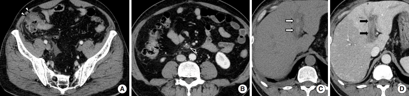
3. 외상과 관련한 문맥혈전증 portal vein thrombosis after trauma
복부 외상은 문맥혈전증의 드문 원인으로 대개 심한 압착 손상과 관련되어 있다. 외상과 관련하여 가장 흔한 문맥 손상소견은 혈전증이며 그 외 찢김, 파열과 이와 동반된 문맥 주위 혈종이 생길 수 있다(Figs. 3, 4). 외상 후에 복강 내 정맥 혈전증이 생기는 병태생리에는 다양한 기전이 작용할 것으로 여겨진다. 가장 흔하게는 전단력(shear force)에 의한 혈관 내피의 손상과 문맥 주위 혈종에 의한 압박으로 문맥혈이 정체되는 것이 원인이 될 수 있다.

Portal vein thrombosis after traffic accident in 50-year-old man. (A) Contrast-enhanced CT image shows ill-defined linear low density lesion in the liver, suggestive of hepatic laceration (arrowheads). (B) Follow-up CT image acquired after 7 days shows the left portal vein thrombosis (arrow). CT, computed tomography.

Portal vein thrombosis after traffic accident in 48-year-old woman. (A, B) Contrast-enhanced CT image shows multiple linear low density lesions in the left hepatic lobe, suggestive of hepatic laceration (arrows). Patent left portal vein (black arrowhead) is noted on initial CT images. (C, D) Follow-up CT images acquired after 6 days show the left portal vein thrombosis (arrowheads). (E) Unenhanced CT image shows hyperattenuating sign, suggestive of acute thrombosis (black arrow). CT, computed tomography.
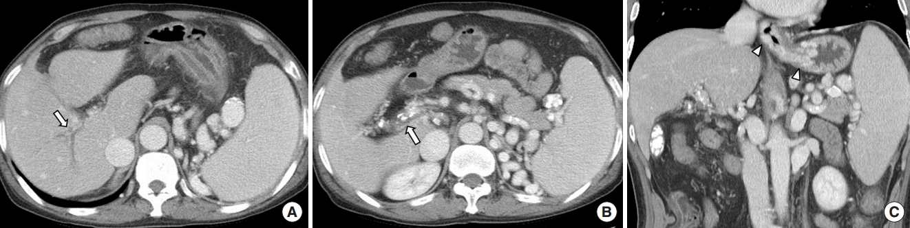
4. 종양성 질환과 관련한 문맥혈전증 portal vein thrombosis associated with neoplasm
Ogren 등[5]은 병원에서 사망한 23,796명의 환자를 대상으로 부검을 하여 문맥혈전증의 평생 유병률을 1.1%로 보고하였다. 문맥혈전증이 있는 환자 중 28%의 환자는 간경화증이 있었고, 23%의 환자는 일차성 간담도암, 44%의 환자는 이차성 간담도 악성종양이 있었으며, 이차성 종양의 경우 췌장암, 위암, 대장직장암, 폐암, 림프종 등의 빈도를 보였다(Fig. 5).

Thrombosis due to a gastric cancer in a 71-year-old man. (A) Contrast-enhanced CT image shows enhancing wall thickening in the stomach, consistent with gastric cancer (arrowheads). (B) CT image shows an expansile thrombus in the left portal vein (arrow). (C) Follow-up image after 3 months shows more increase in extent of the thrombosis (black arrow). (D) Follow-up image after 6 months shows huge hepatic mass (black arrowheads). CT, computed tomography.
간세포암 환자의 6.5-44%에서 간문맥의 종양성 혈전이 보이며 이는 환자의 병기설정과 예후, 치료방침 결정에 주요한 인자로 작용한다[6]. 간세포암이 간문맥을 침범한 경우 대개 종양 재발의 위험이 높고 생존율이 높지 않으므로 수술적 절제, 간이식 같은 공격적인 치료가 부적절한 면이 있다.
종양과 관련하여 문맥혈전증이 발생하는 기전으로는 원발 병소 또는 간으로 전이된 종양에서 종양이 직접 혈관을 침범하여 종양성 혈전이 생길 수 있다. 또한 주변의 비대해진 전이 림프절에 의해 문맥이 압박되거나 부종양 증후군(paraneoplastic syndrome)에 의해 혈액 과응고상태가 일어나면 비종양성 혈전(bland thrombus)이 생길 수 있다[7].
비종양성 혈전은 악성 종양의 유무와 관계없이 생길 수 있으며 만성 간질환 환자의 4.5-26%, 간세포암 환자의 42%에서 보고된다[5]. 한 환자에서 비종양성 혈전과 종양성 혈전이 동시에 존재할 수도 있기 때문에 영상검사에서 문맥혈전증의 발견과 더불어 이 둘을 구분하는 것이 환자의 치료방침 결정에 중요하다. CT 영상에서 간세포암 환자의 문맥혈전이 비종양성 혈전인지 종양성 혈전인지 감별하는 것이 항상 용이한 것은 아니지만 다중시기 조영증강 CT 영상의 동맥기 영상에서 신생혈관(neovascularity)을 동반한 조영증강을 보이고 이환된 혈관 직경이 확장된 경우 종양성 혈전을 의미하며, 이 두 가지 소견이 있을 때 CT의 민감도는 86%, 특이도는 100%로 보고된다(Fig. 6) [6].

Malignant portal vein thrombosis due to a hepatocellular carcinoma in a 65-year-old man. (A) Arterial phase image of contrast-enhanced CT shows ill-defined right hepatic mass, consistent with known hepatocellular carcinoma (arrowheads). Tumor invades the right portal vein (arrow) with avid arterial linear enhancement and expansion of the portal vein. (B) Coronal reformation image shows hypodense tumor thrombus (arrow) is contiguous with a parenchymal hepatocellular carcinoma. CT, computed tomography.
신생혈관은 혈전 내에서 진한 점상 또는 선형의 조영증강을 보이는 것으로 종양성 혈전의 특이적인 소견이다(Fig. 6A). 이 소견은 CT 촬영기법에 따라 민감도에 차이가 있어서 문맥기 영상만을 얻는 단상(single phase) 조영증강 CT에서는 종양성 문맥혈전의 31%에서 신생혈관이 보이나 다중시기 조영증강 CT영상의 동맥기 영상에서는 82%에서 신생혈관이 보이기 때문에 문맥혈전의 감별을 위해서는 동맥기 영상을 포함한 다중시기영상을 얻는 것이 필요하다[6]. 또한 문맥의 확장은 비종양성 혈전에서는 보이지 않으며 이 경우 이환된 혈관 직경은 정상 혈관 직경과 유사하다. 그 외 간실질에 있는 간세포암과 직접적으로 닿아서 침범되어있거나 병변과 연속성을 보이며 이어져 있는 문맥혈전의 경우 종양성 혈전을 의심할 수 있다(Fig. 6B) [6].
최근에는 이중에너지 CT (dual energy CT)를 이용한 material decomposition 방법으로 혈전 내 요오드를 정량화한 후 비교하여 종양성 혈전과 비종양성 혈전을 감별하는 연구들이 이루어지고 있다. 종양성 혈전은 종양세포와 함께 풍부한 신생혈관을 가지는 반면, 비종양성 혈전은 주로 무혈관성 섬유소(fibrin) 또는 피떡(blood clot)으로 구성되어 있다. 혈전의 요오드 밀도는 이론적으로 혈전 내의 혈관분포 정도와 연관되어 있으므로 혈전의 요오드지수를 측정하여 혈전의 특성을 감별하는 데에 이용할 수 있으며 Qian 등[8]은 종양성 혈전이 비종양성 혈전에 비해 유의하게 높은 요오드지수를 보인다고 하였다. 이중에너지 CT를 이용한 혈전 감별방법의 민감도와 특이도는 통상적인 조영증강 CT를 이용한 방법의 그것과 비슷한 정도로 높을 뿐만 아니라 통상적인 CT에서는 평가하기 어려운 작은 혈관의 혈전 발견과 감별에 우월하다고 보고되었다[9,10].
만성 문맥혈전과 관련한 이차적인 형태학적 변화
문맥혈전이 지속되어 만성기가 되면 다양한 형태학적 변화가 동반되어 나타나는데, 간문맥의 해면상변형, 간 내 혈류의 재분포 그리고 문맥전신순환 측부혈관의 발달 등이 있다.
1. 간문맥의 해면상변형 cavernous transformation of the portal vein
간문맥이 막히면 약 1-12개월에 걸쳐서 간문맥 주변으로 작은 측부혈관들이 발달하는데 이를 간문맥의 해면상변형이라 하며, 문맥혈전 내의 재개통과 간문맥 주변의 혈관인 paracholedochal vein 등으로 구성된다. 간문맥의 해면상변형은 간 내 문맥 주변으로도 확장될 수 있기 때문에 간문 주변뿐만 아니라 간 내의 엽상 또는 분절 혈관 주변에도 형성될 수 있다(Fig. 7). 이는 간향성(hepatopetal) 혈류를 보이며 막힌 간문맥 주변으로 내장정맥(splanchnic vein)과 간 내문맥 사이의 우회로 역할을 한다.
Chronic portal vein thrombosis in 60-year-old man. (A, B) Contrast-enhanced computed tomography images show small hypoattenuating portal vein with cavernous transformation at the hepatic hilum and extension around the intrahepatic portal veins (arrows). Calcifications around the portal vein and hypertrophy of the central liver with peripheral atrophic change are also noted, suggestive of chronic change. (C) Coronal reformation image shows splenomegaly and portosystemic collateral veins with gastroesophageal varices (arrowheads), suggestive of the portal hypertension.
간문맥의 해면상변형의 병태생리에 관하여 세 가지 이론이 있는데, 첫 번째는 이 해면종(cavernoma)은 선천성 기형으로서 발달하지 못한 간문맥을 대신하는 구조물이라는 것이고 두 번째는 이를 간문맥의 혈관종으로 생각하는 것이다. 세 번째는 해면종을 문맥혈전증의 결과물로 보는 것이며, de Gaetano 등[11]은 정상 간문맥에 급성 혈전이 생긴 후 전형적인 해면상변형으로 전환되는 9명의 환자를 보고하여 해면종이 문맥혈전증에 대한 반응으로 발생하는 것임을 뒷받침하였다.
간경화증이나 다른 간 질환이 있는 환자에서 문맥혈전증이 상대적으로 흔함에도 불구하고 문맥혈전의 재개통이나 해면상변형은 드물다. 이는 간 질환이 있는 경우 간문맥혈류에 대한 저항이 높아 혈류 속도가 느려지는 것이 원인으로 여겨지고 있으며, 이에 비해 기저 간 질환이 없는 환자들에서 해면상변형의 형성이 더 성공적으로 일어난다고 보고된다[11].
2. 간 내 혈류의 재분포
일반적으로 간 내 혈류에 대한 저항은 질환이 이환된 정도에 따라 부분적으로 불균등하게 나타난다. 혈류에 대한 저항이 올라갈수록 심하게 이환된 부분으로 가는 혈류는 역전되거나 상대적으로 건강한 부분으로 단락을 형성하게 된다. 간문맥의 해면상변형이 간 내로 확장되면 상대적으로 저항이 심한 부분으로 들어가는 문맥혈류는 저항이 적은 다른 분절 정맥으로 혈류가 전환되어 흐르게 되고, 간 내에 문맥혈류의 불균형을 초래한다.
이때 미상엽과 좌간 내 분절을 포함한 간의 중심부는 비교적 문맥혈 공급이 유지되지만 우간의 바깥쪽과 좌간 외 분절은 문맥혈류 부족이 일어난다. 문맥혈이 감소된 부위에 보상적으로 동맥혈류가 증가하므로 조영제의 농도가 높은 동맥혈이 많은 간의 바깥쪽은 더 강한 조영증강을 보일 수 있다. 문맥은 간영양성(hepatotrophic) 혈액을 공급하므로 이러한 혈류의 재분포가 지속되면 문맥혈이 부족한 부위는 만성적인 기능성 경색으로 인해 간세포의 위축이 초래된다. 따라서 간의 중심부는 비교적 문맥혈류가 유지되어 비대해지고, 간의 주변부는 문맥혈류가 감소하여 위축되는 형태학적 변화가 나타난다(Fig. 7A, B).
문맥류
대개의 정맥류는 오금정맥, 목정맥 또는 복재정맥에서 주로 나타나며, 문맥류는 전체 정맥류의 3%로 드물다[1]. 문맥류는 splenomesenteric venous confluence, 주 문맥의 좌, 우 문맥 분지부, 간 내 문맥의 분지부 등 주로 분지부와 합류부에서 호발하며 드물게 비장정맥, 장간막정맥, 제정맥(umbilical vein)에서 나타날 수 있다[1].
문맥류의 원인에 대해 다양한 가설이 제시되는데, 선천적 요인으로 추정하는 이유에는 자궁 내 태아에서도 진단되는 점, 조직학적으로 정상 간에서도 발생하는 점 그리고 추적검사 시 대개 변화가 없는 점 등이 있다. 후천적 요인으로 추정하는 경우는 문맥 고혈압이 있는 환자나 괴사성 췌장염, 이전에 복강 외상이나 수술을 받았던 기왕력이 있는 환자에서 문맥류가 흔히 보임을 이유로 든다[1].
간문맥의 직경은 정상인과 간경화증 환자 모두에서 다양하게 보일 수 있으나, 간 외 문맥의 최대 직경이 정상인에서는 15 mm, 간경화증 환자에서는 19 mm를 넘지 않는다고 알려져 있다. 따라서 국소적으로 주머니모양(saccular) 또는 방추형(fusiform)이면서 간 외 문맥의 경우 20 mm보다 큰 직경을 보이며 나머지 혈관의 직경보다 유의하게 큰 경우 문맥류라고 진단할 수 있다(Figs. 8, 9) [12]. 간 내 문맥의 경우 통상적으로 인정되는 정상 직경의 상한치가 없지만 9 mm보다 큰 경우 문맥류를 의심할 수 있다[12].

Aneurysm of the portal venous system in 69-year-old man with liver cirrhosis. (A) Contrast enhanced CT image shows aneurysmal dilatation of the extrahepatic portal vein with 28 mm in diameter (arrow). (B) CT images show liver cirrhosis, splenomegaly, recanalization of paraumbilical vein, suggestive of portal hypertension. Right portal vein thrombosis with cavernous transformation (arrowhead) is also noted. CT, computed tomography.

Aneurysm of the portal venous system in 78-year-old woman. (A) Contrast-enhanced computed tomography image shows aneurysmal dilatation of the extrahepatic portal vein with 27 mm in diameter (arrow). (B) Curved multiplanar reformation image shows the same findings (arrow).
이는 간 질환이 있는 경우에도 보일 수 있으나, 대다수는 문맥 고혈압이나 만성 간질환이 없는 환자에서 나타나므로 문맥 고혈압이 문맥류 발생에 필수적 인자는 아니다. 문맥류가 있는 환자들은 이와 관련된 증상이 없는 경우가 대부분이지만 혈전이 동반되거나 크기가 크거나 다수의 문맥류가 있는 경우 반복적인 상복부 통증, 황달, 드물게 위장관 출혈 등의 증상이 있을 수 있다[12]. 치료가 필요한 문맥류는 드물지만 30%의 환자에서 혈전증이 발생한다고 보고되며 이 경우 항응고치료가 권장되므로 추적관찰이 권고된다[12].
결 론
간문맥계의 이상소견에 대한 이해는 복강 내 영상소견에 대한 정확한 분석에 도움을 주고 질환의 이해도를 높이는 데에 중요한 정보를 준다. 특히 다중시기 조영증강 CT영상은 간문맥계의 해부학과 관류 이상을 파악하는 데에 용이하고, 문맥혈전증의 경우 종양성 혈전과 비종양성 혈전의 감별에 도움을 준다.