Dislodgement of Two Stents in One Patient during Percutaneous Coronary Intervention
Article information
Abstract
Coronary stent loss is a rare but serious complication during interventional cardiology. This complication occurs not only in the intracoronary area but also in the extracoronary area, such as the aortic root or the left ventricle. An 83-year-old man with stable angina had a stent inserted into a heavy calcific left anterior descending artery. The stent was lost twice during the procedure. The first stent was dislodged from the left main coronary artery to the proximal left anterior descending artery, and the second stent migrated to the aortic root following separation from the balloon. We successfully redeployed the first stent at the dislodged site and retrieved the second stent using a goose-neck snare after moving the stent to the descending aorta. These steps circumvented the need for the patient to undergo emergency cardiovascular surgery.
INTRODUCTION
The incidence of stent loss during percutaneous coronary intervention (PCI) has decreased [1]. However, according to a recent report, stent loss still occurs in 0.32% of cases due to the anatomy of the coronary artery, type of stent, stenting method, and skill of the operator [2,3]. The usual retrieval methods are the small balloon technique, the snare removal technique, and stent deployment [2,3]. Here, we report successful retrieval of two stents using two different methods in one patient. One stent was deployed using a balloon at the dislodged site, and the other stent was retrieved using a goose-neck snare after moving the stent from the aortic root to the descending thoracic aorta.
CASE REPORT
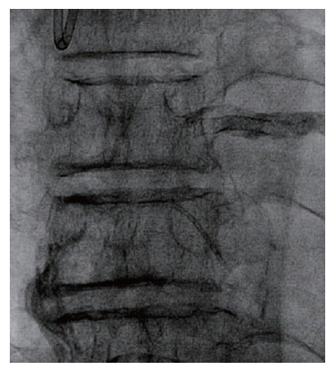
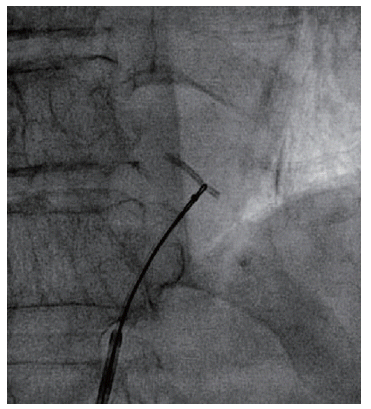
An 83-year-old man was admitted to Soonchunhyang University Gumi Hospital for a herniated intervertebral disc (HIVD) operation. He was referred to our department for a preoperative heart evaluation. His medical history included stable angina and hypertension. Initial echocardiography showed normal left ventricular systolic function and no abnormal regional wall motion. Due to the patient’s medical history and the presence of intermittent chest discomfort, a coronary angiogram was performed. The initial coronary angiogram revealed diffuse, severe stenosis, with acute angulation and heavy calcification of the left main (LM) coronary artery to the distal left anterior descending artery (LAD) (Fig. 1A). The mid-LAD was thought to be the culprit. The right coronary artery also showed mild and diffuse stenosis, with heavy calcification (Fig. 1B). We decided to treat the LAD lesion. An Amplatz (AL1) guiding catheter (7F; Cordis Co., Miami, FL, USA) was used, and the LAD was successfully wired with a balanced middle-weight balloon (Guidant, Indianapolis, IN, USA). After pre-dilatation of the proximal to mid-LAD with 2.5 mm and 3.0 mm Sprinter Legend balloons (Medtronic, Minneapolis, MN, USA), a 3.0×23 mm Genous (OrbusNeich, Hong Kong;drug-eluting [sirolimus]anti-CD 34 monoclonal antibody-coated) stent was planned to be deployed at the lesion to reduce the risk of restenosis and the duration of dual antiplatelet therapy. However, it proved difficult to advance the stent through the tightest lesion due to the acute angle and heavy calcification. Thus, we placed the stent at the proximal lesion for additional back-up before stenting the mid-LAD lesion using the deep-seating method. After stenting at the proximal lesion, repeated ballooning of the tightest mid-LAD lesion was performed. During deployment of the second 3.0×23 mm Genous (drug-eluting[sirolimus]anti-CD 34 monoclonal antibody-coated) stent, it became stuck and was dislodged due to the proximal calcification of the LAD. The stent that had been deployed earlier was also dislodged (Fig. 2). We attempted to expel the stent to avoid embolization using two parallel wires. When this failed, we passed a low-profile balloon distal to the dislodged stent and tried to pull back the balloon to retrieve the stent. However, this attempt also failed. When then tried to deploy the stent at the proximal lesion and inflate the dislodged stent fully using a large-sized balloon. We attempted to deploy a shorter 3.0×18 mm Genous stent to treat the culprit lesion. At that time, the patient was scheduled to undergo surgery for severe HIVD-related symptoms uncontrolled by medication and rehabilitation. Therefore, we selected a Genous stent, which requires a short period of dual anti-platelet therapy. This type of stent is also associated with a decreased risk of restenosis, and there is no difference in the rate of stent thrombosis and other clinical outcomes compared to other types of stents. During deployment of the third stent, the stent was dislodged in the LM at the prior site of the stent. We pulled the stent backward, but it came free from the balloon catheter and migrated to the aortic root (Fig. 3). We tried to grab and remove the stent using a goose-neck snare at the aortic root level, but this failed. The stent was very difficult to grab because the distal part was rotated, and the proximal part was fixed at the aortic valve (AV). Thus, we touched the stent using 0.8636-mm Terumo wire, which was fixed at the AV structure. As the aortic arch angle of the patient was very obtuse, we assumed that the stent would migrate to the peripheral rather than the cranial level if we touched and stimulated the stent continuously. We used Terumo wire to move the stent to the peripheral level. Fortunately, the stent migrated from the aortic root into the descending thoracic aorta (Fig. 4). We then removed the stent successfully using 10-mm a goose-neck snare (Fig. 5). The patient remained hemodynamically stable, without chest pain. Angiography showed good thrombolysis in myocardial infarction flow. We attempted a second PCI but stopped the procedure after along duration at the patient’s insistence.

(A) Baseline coronary angiography shows diffuse severe stenosis with acute angulation and heavy calcification of the left main coronary artery to the distal left anterior descending artery. (B) The right coronary artery also had mild and diffuse stenosis, with heavy calcification.

The stent was dislodged at the distal main and proximal left anterior descending artery (arrow).
The third stent migrated to the aortic root during deployment.

Moving the stent (arrow) from the aortic root to the descending thoracic aorta using a 0.8636-mm Terumo wire.
Removing the stent successfully using a goose-neck snare at the descending thoracic aorta level.
DISCUSSION
The incidence of stent loss during PCI has decreased due to improvements in stent technology and operator’s skills [1]. The small balloon technique is easy, and the success rate is high [3]. The snare removal technique is also a common method, with a high success rate [2]. In the latter, a snare is used to capture the stent and pull it into the guiding catheter.
In our case, when the first stent was lost, we made three attempts to avoid stent embolization using both of these techniques with a parallel wire technique. All the retrieval methods failed because the stent was stuck in the heavily calcified LAD artery. The structure of the lesion at the proximal LAD also impeded the retrieval. When stent retrieval is difficult, crushing or deploying the stent in a nontarget lesion is another option. This technique can reduce the contrast load and decrease the procedure time, as well as avoid stent embolization [4]. We decided to deploy the stent at the proximal lesion and inflate the dislodged stent fully using a large-sized balloon. A follow-up angiogram showed good flow patency.
Stent loss can be a problem not only in cerebral and peripheral arteries but also in coronary arteries [2,4,5]. A report of stent embolization and retrieval using the two-wire technique at the femoral artery level has been published [6]. In our case, we tried to remove the stent at the aortic level. The distal part of the stent was rotated because of forward aortic blood flow, and the proximal part was fixed at the AV structure, which made it difficult to grab the stent using the goose-neck snare. Due to the obtuse angle of the aortic arch, a stent may move to the peripheral vasculature of, for example, the limbs, than intracranial arteries. We removed the stent successfully using a 10-mm goose-neck snare after moving the stent from the aortic root to the descending thoracic aorta [7].
It is important to avoid embolization and surgery. When a stent is lost in an extracoronary area, moving the stent to a safe area is an alternative method to avoid embolization and surgery.
In conclusion, stent dislodgment, loss, and embolization can occur during PCI. Our meticulous attitude and advanced stent technique need. We described an interesting and instructive case of the dislodgement of two stents in one person during coronary angioplasty and the retrieval of the stents using various retrieval methods without surgery.