INTRODUCTION
The Hymenoptera order is divided into three families: Apids, Vespidae, and Formicidae. Apids include the honeybee, bumblebee, and sweat bee, which are all docile and tend to sting mostly on provocation [1]. The yellow jacket, yellow hornet, white (bald)-faced hornet, and paper wasp all belong to the Vespidae family. The Formicidae family includes the harvester ant and the fire ant.
When a ‘bee’ sting results in a large local reaction, defined as >5 hours in and lasting >24 hours, the likelihood of anaphylaxis from a future sting is about 5%. Reactions to Hymenoptera stings [1–5] are classified into normal local reactions, large local reactions, systemic anaphylactic reactions, systemic toxic reactions, and unusual reactions. The most frequent clinical patterns are large local and systemic anaphylactic reactions.
Bee venom acupuncture (BVA) involves injecting purified and diluted bee venom into acupoints [6]. BVA exhibits several pharmacological actions, including analgesic, anti-inflammatory, anti-arthritic, and anti-cancer effects through multiple mechanisms, such as activation of the central inhibitory and excitatory systems and modulation of the immune system [7]. The analgesic effects of BVA have been reported in animal experiments [8] and in the clinic [9].
CASE REPORT
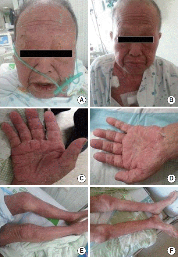
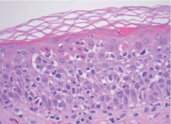
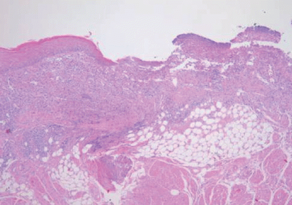
A previously healthy 75-year-old man visited Soonchunhyang University Bucheon Hospital because of poor appetite due to oral ulcer. He had skin eruption, oral ulcer, urinary retention, and conjunctival itching. But he had no other remarkable medical history including allergy. On physical examination late inspiratory crackles in both lower lung fields auscultated. Generalized prutirtic skin eruption showed in whole body. He had pruritic acute urticaria after acupuncture and then 2–6 weeks this skin lesion developed (Fig. 1A, C, E). The blood pressure was 125/75 mm Hg, the pulse 65 beats per minute, the oral temperature 36.5°C, the respiratory rate 18 breaths per minute, the oxygen saturation 95.7%, pH 7.447, PaO2 74.7 mm Hg, and PaCO2 28.1 mm Hg while the patient was breathing ambient air. An electrocardiogram revealed normal sinus rhythm. Pulmonary function test revealed forced expiratory volume in one second (FEV1)/forced vital capacity (FVC) 97%, FVC 1.28 L (41%), FEV1 1.32 L (60%), and diffusing capacity for carbon monoxide 15.1 mL/mm Hg (71%) indicating restrictive pattern. Chest radiographs revealed bilateral patchy opacities, more in the right lung than in the left lung. Computed tomography of the chest revealed multifocal consolidation and ground-glass opacities in all lobes of both lungs. Computed tomography of the abdomen revealed small and large bowel was dilated including air and lots of fluid, likely infectious enterocolitis. Total immunoglobulin E (IgE) was 623 kU/L. The patient underwent skin-prick testing 14 days after the event. Specific IgE for Dermatophagoides farinae, Dermatophagoides pteronyssinus, cat fur, dog fur, mugwort, and ragweed were negative. We carried out bronchoscopy and the result was no endobracheal lesion. Bronchoalveolar lavage revealed machrophage 17.20%, neutrophil 71.60%, and eosinophil 11.20%. We have considered the possibility of Stevens-Johnson syndrome following 5 times BVA history and he had whole body skin eruption, oral ulcer, and interstital pneumonia. Punch biopsies were taken from the skin lesion on chest and tongue. Microscopically, the skin biopsy specimen showed orthokeratotic hyperkeratosis, superficial dermal lymphocytic infiltration with lymphocytic exocytosis, and scattered necrotic keratinocytes (Fig. 2). Biopsy of the tongue lesion showed severe ulceration of surface epithelium with inflammed granulation tissue (Fig. 3).
The patients were diagnosed as Stevens-Johnson syndrome and treated with systemic steroid and anti-histamine. Lots of skin lesions were subsided and angioedema was disappeared (Fig. 1B, D, F). Redness was subsided and desquamation was sustained for 2 weeks. He was informed of the results and of the risk for bee venom in the future.
DISCUSSION
Venom hypersensitivity [1–5], as defined in the recently revised nomenclature for allergy, may be mediated by immunologic mechanisms (IgE-mediated or non-IgE-mediated venom allergy) but also by nonimmunologic mechanisms.
Venom hypersensitivity diagnosis [5] should be collected on: number and date of sting reactions, sort and severity of symptoms, interval between sting and the onset of symptoms, emergency treatment, sting site, retained or removed stinger, environment and activities before sting, risk factors of a particular severe reaction, risk factors for repeated re-stings, tolerated stings after the first systemic reactions, and other allergies.
The skin, the gastrointestinal, respiratory, and cardiovascular systems can be involved. Severe reactions or a status after resuscitation may leave patients with a permanent disorder: hypoxic brain damage with permanent neurologic deficits, and myocardial infarction [5]. For comparison, when there is a history of anaphylaxis from a previous Hymenoptera sting and the patient has positive skin tests to venom, at least 60% of adults and 20% to 32% of children will develop anaphylaxis with a future sting [1–4]. Both patient groups should be instructed about avoidance measures and carrying and knowing when to self-inject epinephrine, but immunotherapy with Hymenoptera venom is indicated for those patients with a history of anaphylaxis from the index sting and not for patients who have experienced a large local reaction [1].
Bee venom acupuncture has been used in Korea to relieve various pain symptoms. Bong-Chim therapy [10] can be used for various diseases. Bong and Chim are Sino-Korean words meaning bee and stinger or acupuncture. There are two modalities in Bong-Chim therapy. One is live bee acupuncture, which applies honeybee (Apis cerana) stingers directly into the lesion, and the other is injection of extracted bee venom into the lesion with a syringe. The former is a traditional and popular modality, whereas the latter has only recently been attempted. Many people who have received live bee acupuncture therapy have reported skin symptoms, which are called Bong-Chim dermatitis, and more precisely defined as live bee acupuncture dermatitis. The acute stage is an inflammatory reaction, such as anaphylaxis or urticaria. In the chronic stage, a foreign body granuloma may develop from the remaining stingers, similar to that of a bee sting reaction. The subacute phase, the characteristic histological flame figures as a characteristic feature of live bee acupuncture dermatitis that is different from the adverse skin reaction of accidental bee sting [10]. In this case acute and subacute stage, the characteristic urticaria, angioedema and Steven Johnson syndrome show as a characteristic feature of live bee acupuncture dermatitis that is different from previous report [7].


