INTRODUCTION
Percutaneous endoscopic gastrostomy (PEG) was first used in clinical practice in 1980 and has become an established method for providing enteral nutrition in a diverse range of neurological and malignant conditions to patients who are unable to swallow or maintain adequate nutrition [1]. PEG is associated with a number of immediate and delayed complications. Typically, buried bumper syndrome (BBS) is a late consequence occurring 3–6 months after PEG tube placement [2]. The condition is often a result of excessive pressure exerted on the gastric mucosa by the tight anchorage and subsequent excessive tension between the internal and external bumpers leading to mucosal erosion and embedding of the internal bumper in the gastric wall, causing feeding obstruction [3,4]. Few documented reports of acute BBS occurring as early as 14 days after PEG tube placement have been published in literature [2,5]. Herein we report two cases of early onset BBS, successfully managed with endoscopic treatment, together with an update review of the English or Korean literature.
CASE REPORTS
1. Case 1: Early onset buried bumper syndrome with apparent abscess of the abdominal wall
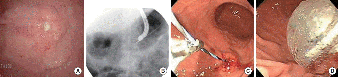
A 78-year-old male was admitted to division of gastroenterology due to melena, abdominal discomfort and fever that had developed 2 days prior. The patient had a medical history of left middle cerebral artery infarction. He had suffered from recurrent aspiration pneumonia and thus underwent PEG tube insertion using Ponsky PEG Kit (Bard Endoscopic Technologies, Billerica, MA, USA) 8 days before admission. At that time, the PEG tube was well-positioned in the gastric lower body lessor curvature without any complication. The body temperature was 37.8°C, the pulse was 88 beats/min, respiration was 22/min, and blood pressure was 160/90 mm Hg. Laboratory results showed the white blood cell count was 23,100/μL, neutrophil 84%, hemoglobin 5.9 g/dL, platelet count 433,000/μL, erythrocyte sedimentation rate 114 mm/hr, and C-reactive protein 8.76 mg/dL. Upper endoscopy was performed to evaluate the cause of melena and showed deep ulceration with exudates surrounding the internal bumper and embedding of the internal bumper into the gastric wall (Fig. 1A). Computed tomography (CT) revealed an ill-defined enhancing lesion with fat infiltration and air bubbles around the gastrostomy track (Fig. 1B). The patient was diagnosed with upper gastrointestinal bleeding and abdominal abscess due to acute BBS. Therefore, we immediately removed the buried PEG tube with external traction using minimal exertion and administered broad spectrum antibiotics to allow the original site to heal before attempting replacement of the PEG tube. A high-dose proton pump inhibitor was also used to decrease gastric secretions and prevent rebleeding. A Foley catheter was inserted 3 days after PEG tube removal to maintain the old track site. The abscess around the track was nearly resolved 27 days after PEG tube removal (Fig. 1C). The new PEG tube was reinserted through the old track after removal of the Foley catheter under endoscopic guidance (Fig. 1D).
2. Case 2: Early onset buried bumper syndrome without apparent abscess
A 67-year-old male developed double hemiplegia, dysphagia and aspiration of food during meals as a result of a multiple territory infarction. Therefore using Ponsky PEG Kit (Bard Endoscopic Technologies), a PEG tube was placed on the mid-body greater curvature for enteral feeding. No complication related to the procedure developed at that time. Eleven days post-procedure, the patient presented to our hospital complaining of abdominal pain and an inability to infuse food through the tube. With the exception of a 38.0°C fever, his vital signs and laboratory tests were unremarkable. The patient subsequently underwent a CT scan of the abdomen that revealed the internal button of the gastrostomy tube in the abdominal wall layer without any evidence of abscess or peritonitis. Upper endoscopy also showed an ulcer-like mucosal defect in the previous internal opening site of the PEG tube (Fig. 2A). The patient was diagnosed with acute BBS without abscess. The old PEG tube was removed manually with external traction using minimal exertion and the zig-zag guidewire was inserted through the previous track under fluoroscopic guidance (Fig. 2B). A balloon-type gastrostomy tube was successfully placed over the guidewire, with the guidewire tightly fixed using grasping forceps (Fig. 2C, D). The patient was given antibiotic treatment for 3 days after the procedure. No complication developed related to the procedure. Subsequently, his wound care was completed and he started enteral feeding via the PEG tube during hospitalization; he was discharged to the nursing home in stable condition.
DISCUSSION
BBS was first described in 1988 and the reported incidence in the majority of the literature is typically 0.3% to 8% [6]. Typically, BBS is considered a long term consequence of tight apposition of the external bolster of the gastrostomy tube slowly erodes into the gastric wall as tension is created on the gastrostomy tube tract, which causes pain and the inability to infuse feeding [7]. The earliest reported case due to complication was at 8 days post-insertion. All types of PEG feeding tubes (initial placement, replacement tubes, and low profile devices) are associated with this complication [8], as observed in our patient cases with acute BBS. One patient was diagnosed with BBS at 8 days post-insertion and the other patient at 11 days post-insertion.
Although this is not a frequent complication (250/100,000 PEG patients per year), every interventional endoscopist should be able to diagnose and treat this condition. Radiological contrast studies are insufficient for the diagnosis of BBS; upper gastrointestinal endoscopy is mandatory in all cases of PEG tube malfunction.
There are optional procedures for BBS that restore PEG tube function using endoscopic techniques. The treatment of BBS depends on the type of PEG tube. If the internal bolster is collapsible, as it is on externally removable PEG tubes, the PEG tube can be removed by simple external traction. In a modification of this technique, the PEG tube can be cut short and a guidewire passed through the stump into the gastric cavity. The guidewire is snared endoscopically and pulled out of the oral cavity and attached to a new PEG tube. The guidewire at the abdominal surface is pulled, pulling the new PEG tube into the gastric cavity. The dilating portion of the new PEG tube engages the buried bumper on the old PEG tube. As the new PEG tube is pulled through the abdominal wall, the old PEG tube is pushed out of the abdominal wall and removed [2].
However, if the internal bumper on the PEG tube is rigid, the PEG tube may need to be removed by PEG wound tract cut-down or the push-pull T-technique. The push-pull T-technique requires the PEG tube to be cut 3 cm from the abdominal wall. An endoscope is introduced into the stomach, and a snare is passed through the scope and through the PEG tube opening in the gastric wall. Once the snare is protruding externally through the PEG tube, an additional short piece of PEG tube is cut from the excess PEG tubing. The snare is opened, and this short piece of tubing is grasped and pulled back against the PEG tube creating a T-shape. A Kelly clamp is placed across the T-shape. The endoscopist slowly removes the endoscope, snare, and PEG tube orally as a second operator pushes the Kelly clamp and PEG tube into the gastric lumen. This combined procedure frees the internal bumper from the gastric wall. Once the PEG tube is removed, a new PEG tube can be placed through the existing PEG tract using direct endoscopic visualization. A standard PEG tube placement technique should be used to permit the PEG tube dilator to re-expand the partially closed PEG tube tract [3].
Patients presenting with a BBS are usually in poor general condition and often not suitable for general anesthesia. In that case, the endoscopic procedure, performed under conscious sedation, offers a much better option than surgical removal in these patients [9]. However, the endoscopic approach requires special expertise, is not always successful, and may carry the risk of bleeding or perforation. Although the endoscopic approach may be the first choice in patients unfit for general anesthesia, laparoscopy should be considered where endoscopy has failed or is not possible as in patients with head and neck cancers who cannot be intubated for endoscopy [10].
In conclusion, a PEG tube in a patient with early onset BBS can be safely removed endoscopically without a skin incision or gastric wall disruption. A simple and safe endoscopic removal technique was described in the present case report.