INTRODUCTION
Technetium-99m methoxyisobutrlisonitrile (Tc-99m MIBI) scan has markedly improved clinical decisions to localize hyperfunctioning parathyroid tissue before operation [1]. Adenomatous and hyperplastic parathyroid tissue show higher uptake and lesser washout of Tc-99m MIBI than adjacent thyroid tissue. Radioiodine scan is useful to differentiate thyroid lesion and hyperfunctioning parathyroid tissue. Also, Radioiodine scan may facilitate accurate imaging diagnosis in several thyroid lesions that accumulate MIBI [2]. This case underlines the need of non-invasive nuclear imaging for preoperative work up of secondary hyperparathyroidism.
CASE REPORT
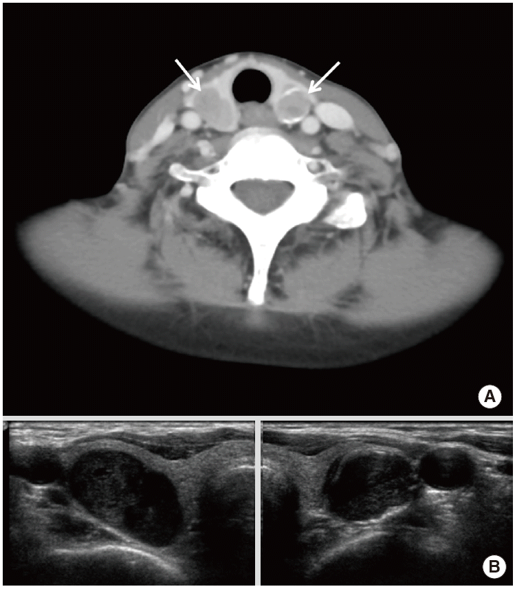
A 47-year-old female presented with incidental thyroid nodule and elevated serum parathyroid hormone (PTH) level. She had reported no history of pruritus or psychiatric symptom. Her medical history includes a 17-year medical management of hypertension and a 15-year history of hemodialysis for chronic renal failure resulting from immunoglobulin A nephropathy. Initial physical examination revealed blood pressure was 145/79 mm Hg, palpable mass in anterior neck without other abnormality. Contrast-enhanced computed tomography (CT) of the neck showed poorly enhanced and well defined oval shape nodules in both thyroid lobes (Fig. 1A). Subsequent ultrasonography (USG) demonstrated two circumscribed oval hypoechoic nodules in the both thyroid lobes (Fig. 1B). Blood tests revealed increased serum levels of intact PTH (greater than 3,000 pg/mL; normal range, 15.0 to 68.3 pg/mL) and serum phosphorus (6.9 mg/dL; normal range, 2.5 to 5.5 mg/dL), with normal calcium level (10.4 mg/dL; normal range, 8.0 to 10.5 mg/dL). These findings were suggestive of secondary hyperparathyroidism and surgical approach was considered following the extremely elevated levels of PTH.
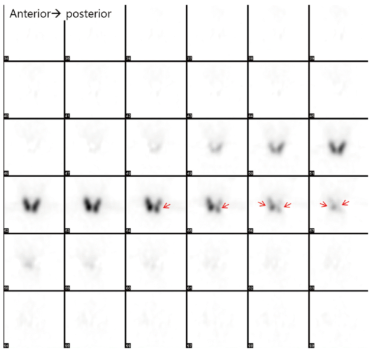
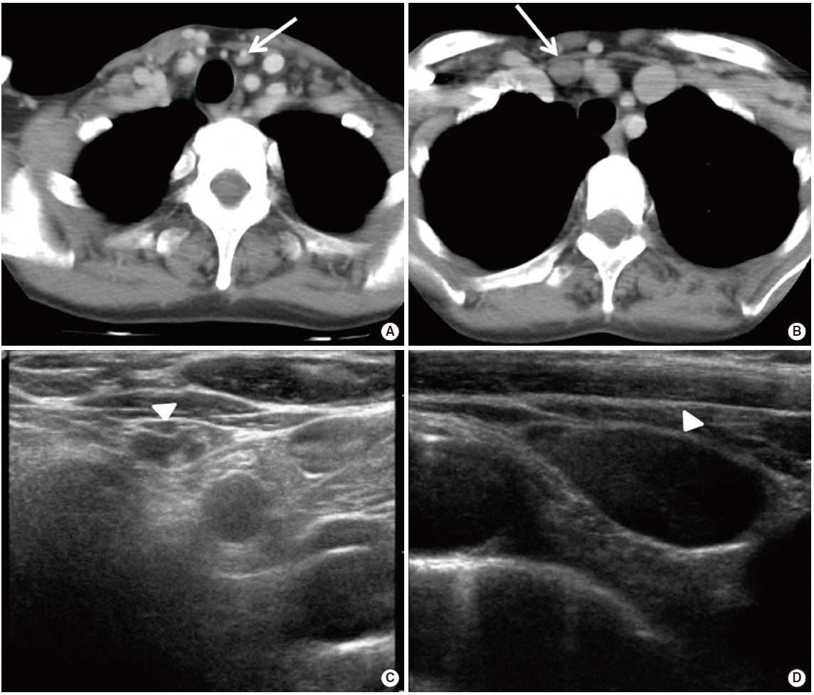
Preoperative dual phase Tc-99m MIBI scan was performed at 15 and 120 minutes after injection of 740 MBq Tc-99m MIBI. Each image was obtained by a dual headed gamma camera (Infinia GP; GE Healthcare, Milwaukee, WI, USA) equipped with a low energy general purpose collimator. Delayed images were obtained after 2 hours and showed 4 focal Tc-99m MIBI uptakes. Of these, two uptake foci were located at intrathyroid area, and the remaining two were around the lower pole of left thyroid lobe and right upper mediastinal area (Fig. 2). To differentiate false positive finding from thyroid gland origin, I-123 scan and single photon emission tomography (SPECT) were performed 24 hours after injection of 111 MBq of I-123. On I-123 SPECT, only the normal thyroid gland uptake was shown and no definite abnormal uptake was noted around the thyroid gland. The coronal view of I-123 SPECT showed photon defect in the thyroid gland, corresponding to the two foci of focal Tc-99m MIBI uptake at the intrathyroidal lesions (Fig. 3). CT and USG images were retrospectively reviewed and nodular lesions were found in the right supraclavicular fossa and left neck level VI corresponding to the findings of radionuclide studies (Fig. 4).
Subsequently, the patient underwent bilateral intrathyroidal mass nodule enucleation with perithyroidal and mediastinal nodule excision. Histopathological results of postoperative specimens revealed parathyroid hyperplasia. Post-surgery, the serum PTH and phosphorus levels decreased to normal ranges (20.7 pg/mL and 2.2 mg/dL, respectively).
DISCUSSION
Chronic renal failure results in hyperphosphatemia with subsequent of calcitriol decrease fromalpha hydroxylation of cholecalciferol. The resulting hypocalcemia stimulates PTH secretion (secondary hyperparathyroidism) increases parathyroid gland (hyperplasia) and is common in chronic renal failure patient [3]. Salem [4] reported that more than 50% of patients receiving long term hemodialysis showed more than three times higher PTH level than normal.
Medical management of secondary hyperparathyroidism decreases the need for surgical intervention of parathyroid gland lesions. Indications of surgical intervention include markedly elevated PTH levels with one of followings: symptoms (bone and joint pain, muscle weakness, pruritus, psychic irritability, etc.), metabolic bone disease, or intact PTH concentration greater than 800 pg/mL associated with hypercalcemia and/or hyperphosphatemia [5].
The patient in our case report showed extremely elevated serum PTH level (more than 3,000 pg/mL), hence, the patient was treated with surgical approach. The success of surgical approach is highly dependent on precise preoperative localization of pathologic parathyroid lesions. The most common reason for persistent hyperparathyroidism is failure to remove all hyperfunctioning parathyroid tissue. Tc-99m MIBI scan is a useful technique for localization of hyperfunctioning parathyroid tissue in primary parathyroid hyperparathyroidism. Sensitivity of more than 90% has been reported in localizing single parathyroid adenoma [2,6]. However, previous research has reported lower sensitivity for parathyroid hyperplasia in patients with secondary or tertiary hyperparathyroidism [7,8]. Tc-99m MIBI scan in our case found two parathyroid lesions with intrathyroidal ectopic parathyroid gland locations. Although there is still no consensus of the role of Tc-99m MIBI scan in secondary or tertiary hyperparathyroidism, Tc-99m MIBI scan provided valuable information in our case.
Simultaneous use of Tc-99m MIBI and I-123 scan can improve the imaging of parathyroid glands in secondary hyperparathyroidism [9]. This patient showed two intrathyroidal and two ectopic thyroidal lesions on delayed Tc-99m MIBI scan. I-123 is considered to be the optimal thyroid agent for dual-tracer parathyroid imaging. It was performed alongside SPECT to differentiate the thyroid lesions. Two intrathyrodal lesions showed lesser uptake of I-123 than surrounding normal thyroid tissue, while 2 ectopic lesions did not show I-123 uptake, therefore suggesting that all four nodular lesions originated from extra-thyroidal tissue.
Using a pinhole collimator can significantly improve the sensitivity of the parathyroid scan [10,11]. SPECT provides improvement of sensitivity in comparison to planar imaging for localization and differentiation of thyroid lesions from parathyroid lesions [12,13]. We also performed I-123 SPECT for characterization of nodular lesions. Hybrid SPECT/CT might help ascertain accurate preoperative work-up. Intraoperative use of gamma probe for localization of parathyroid glands is also useful methods [3,14].
The uptake of Tc-99m MIBI in primary hyperparathyroidism might be influenced by a variety of biochemical factors [15,16]. The decreased gradient of Ca level in secondary hyperparathyroidism between pre- and post- operative serum test may correlate with the positive finding of Tc-99m MIBI scan and high PTH level.
In the present case, all 4 lesions including 2 intrathyroidal and 2 ectopic hyperplastic parathyroid gland lesions were accurately localized using Tc-99m MIBI and I-123 scans in patient with secondary hyperparathyroidism. This suggests radionuclide scan may have a wider role in the surgical management of secondary or tertiary hyperparathyroidism.


