SMS 2011 December;17(2):112-114.
Published online 2011 December 30 |
| Copyright ⓒ 2010 Soonchunhyang Medical Science
|
| A Case of Trauma-Induced Single Xanthelasma Palpebrarum |
| You In Bae, Ji Hoon Sim, Jung Hoon Yang, Sanghoon Lee, Young Lip Park |
| Department of Dermatology, Soonchunhyang University Bucheon Hospital, Soonchunhyang University College of Medicine, Bucheon, Korea |
| Corresponding Author: Young Lip Park , Tel: +82-32-621-5062 , Fax: +82-32-621-5018 , Email: ylpark@schmc.ac.kr
|

|
ABSTRACT
|

|
|
| A 33-year old Korean man visited our department complaining of single mildly pruritic yellowish flat-topped papule on left upper eyelid. Lipid profile including low density lipoprotein-cholesterol and triglyceride level was above normal range. He remembered that the site was scalded by boiling oil about eight months ago. The patient was diagnosed with xanthelasma palpebrarum (XP) based on the clinical and histopathological findings, including diffuse infiltrate of large, pale-staining cells which have abundant foamy cytoplasm in the dermis. He was treated with CO2 laser ablation followed by application of 70% trichloroacetic acid with wooden stick. After a month, the skin lesion showed moderate improvement with disappearance of yellowish color and flattopped elevation. So far, case of XP developed after minor trauma has not been reported. This case illustrates that minor trauma could be a trigger factor in the development of the disorder. |
|
Keywords: Trauma; Trichloroacetic acid; Xanthelasma palpebrarum |
|
|
|
INTRODUCTION
|
|
|
Xanthelasma palpebrarum (XP) is the most common type of xanthoma. It develops on the eyelids and is usually symmetric
[1]
. This disorder may be accompanied with some familial lipoprotein disorders and systemic hyperlipidemia especially hypercholesterolemia; however, more than half of the patients are normolipemic
[1,2]
. Though a few cases of Koebnerization or trauma induced in eruptive xanthoma have been reported, however, trauma induced XP has not been reported so far except for one case
[3-5]
. Various methods for treatment of xanthelasma include surgical excision,
cauterization with chemical peeling agent, laser ablation, and so on
[6]
. The authors experienced excellent outcome after treatment with combination of CO2 laser ablation and trichloroacetic acid (TCA) cauterization. |
|
|
CASE REPORT
|
|
|
A 33-year-old Korean man visited our department complaining of single mildly pruritic papule on left upper eyelid. He remembered that boiling oil spattered while he was deep-frying about eight months ago. The skin lesion became bulla in several days. Therefore the patient fully believed that it was a scalded scar. He disinfected the wound with alcohol gauze and topical antibiotics by himself. After three to four weeks, he noticed the wound became elevated and subjective symptoms such as mild pruritus developed.
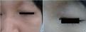
On physical examination, the lesion was about 0.8×0.4 cm sized; showed yellow colored and flat-topped appearance
(Fig. 1A)
. Other similar lesions were not found and he was otherwise healthy. The family history was noncontributory. Laboratory tests including lipid and lipoprotein level were checked. Total cholesterol, low density lipoprotein (LDL)-cholesterol and triglyceride level was 265 mg/dL (90 to 250 mg/dL), 174 mg/dL (<100 mg/dL), and 228 mg/dL (<200 mg/dL), respectively. High density lipoprotein-cholesterol level was within normal range (45 mg/dL).
The patient was given the presumptive diagnosis of hypertrophic scar or xanthelasma. To determine nature of the skin lesion, we performed a 3 mm punch biopsy. Histopathology showed flattened epidermis with mild hyperkeratosis. The dermis was diffusely infiltrated by large, foamy cells and histiocytes (Fig. 2A). A few giant cells were also found (Fig. 2B). Consequently, he was diagnosed with xanthelasma.
Since he wanted to remove the lesion, we decided to perform laser ablation followed by TCA application. The procedure was performed after occlusion with local anesthetic cream (eutectic mixture of local anaesthetics). First, the lesion was concavely ablated by the CO2 laser until some white dermal components were exposed. Grossly, when about 70 to 80% of the yellow materials were removed, 70% TCA was applied with wooden stick. After application of topical antibiotic ointment (mupirocin), the wound was occluded by hydrocolloid dressing (DuoDERM Extrathin, ConvaTec International, Skillman, NJ, USA). The skin lesion was assessed by two dermatologists at 2, 4, 6 weeks after initial treatment. At 6th week, the lesion showed marked flattening and the yellow color was vanished in most part
(Fig. 1B)
except for the margin of the lesion where ablation was not fully performed. Over six-month follow-up period since the initial treatment, there had been no recurrence and no complication developed. He was also referred to department of endocrinology in our hospital for further evaluation of his lipid abnormalities. He was recommended to get management through dietary modification and exercise. |
|
|
DISCUSSION
|
|
|
Xanthoma is caused by deposition of lipids in large foam cells in the tissue. Types of xanthoma are classified mainly by clinical presentation and site of involvement. Also, various different genetic disorders of lipid metabolism may present with typical xanthoma pattern. XP is a benign skin disorder characterized by yellowish to orange colored papules usually on upper eyelid and is most common type of xanthoma
[1,2]
. It is usually considered to develop more commonly in female than in male
[1]
. This painless disorder frequently develops symmetrically and tends to be permanent and progressive. Some genetic disorders, such as familial hypercholesterolemia, familial dysbetalipoproteinemia, and familial defective apolipoprotein B-100 may be associated with the disease. It may occur in patients with hyperlipidemia, especially hypercholesterolemia. Therefore, patients of XP with a young age of onset and a family history of hyperlipidemia are at higher risk of having lipid abnormalities. However, more than half of the XP patients are normolipemic
[2]
.
While there are a few reported cases of trauma induced or Koebner phenomenon in eruptive xanthoma
[3,4]
, trauma associated XP is seldom reported. In one report, a 47-year-old woman with XP was treated by TCA application and experienced extension of the skin lesion
[5]
. In the case, however, it is not sure whether extension of the disease was merely due to natural progression or a result from Koebnerization. In contrast, our patient did not have any history of XP and had a clear history of minor trauma on the affected site. Though he had no known family history of hyperlipidemia or familial lipoprotein disorders, laboratory profiles including lipid & lipoprotein profile were elevated. Moreover, even though the possibility of spontaneous development of XP cannot be fully excluded, since he had no XP on other part of his body for last a few months of follow up period, we could confirm the lesion as a rare case of XP that resulted from minor trauma.
The pathogenesis or possible mechanism of trauma in the development of XP is not yet understood. Because he had elevated LDL-cholesterol and triglyceride level, we assume that minor trauma evoked a series of inflammation which can trigger or initiate macrophage accumulation, cholesterol or triglyceride uptake, and foam cell formation in the affected area. There is an interesting case of XP following allergic contact dermatitis from para-phenylenediamine in a black eyelash-tinting product
[7]
. In the report, the authors adopted the hypothesis that increased lipid peroxidization may lead to formation of xanthelasma
[8]
.
Treatment of XP is challenging due to its location and tendency to recur. Though the best method is surgical excision, it is not always feasible. Since the patient was not willing to get a surgical removal, we chose rather traditional method which is combination technique with CO2 laser ablation followed by 70% TCA application. After a single session of treatment, yellow colored materials disappeared in most area, and the lesion was replaced by reepithelialized wound. Though longer follow-up visit may be needed, treated lesion showed no recurrence or scarring during 6 months of follow-up period.
In summary, the authors experienced a very rare case of XP that developed after minor trauma and suggest that any trauma may trigger XP especially in a patient with underlying abnormalities in lipid profile. |
|
|
|
FIGURES
|
|
|
|
Fig.1
(A) A single yellow colored flat-topped papule on upper medial eyelid. (B) Six weeks after treatment, xanthelasma lesion was removed and replaced by reepithelizing erythema in most part. |

|

|
Fig.2
(A) Flattened epidermis with mild hyperkeratosis. The dermis was diffusely infiltrated by large, foamy cells and histiocytes (H&E, ×200). (B) A few of giant cells were found (H&E, ×400). |
|
|
|
| |
|
|
REFERENCE
|
|
|
|
1.
|
Bergman R. The pathogenesis and clinical significance of xanthelasma palpebrarum. J Am Acad Dermatol 1994;30(2 Pt 1):236-42. |
|
2.
|
White LE. Xanthomatoses and lipoprotein disorders. In: Wolff K, Goldsmith LA, Katz SI, Gilchrest BA, Paller AS, Leffell DJ, editors. Fitzpatrick’s dermatology in general medicine. 7th ed. New York: McGraw Hill; 2008. p. 1272-80. |
|
3.
|
Goldstein GD. The Koebner response with eruptive xanthomas. J Am Acad Dermatol 1984;10:1064-5. |
|
4.
|
Miwa N, Kanzaki T. The Koebner phenomenon in eruptive xanthoma. J Dermatol 1992;19:48-50. |
|
5.
|
Akhyani M, Daneshpazhooh M, Jafari AK, Naraghi ZS, Farahani F, Toosi S. Koebner phenomenon in xanthelasma after treatment with trichloroacetic acid. Dermatol Online J 2006;12:12. |
|
6.
|
Rohrich RJ, Janis JE, Pownell PH. Xanthelasma palpebrarum: a review and current management principles. Plast Reconstr Surg 2002;110:1310-4. |
|
7.
|
Bhat J, Smith AG. Xanthelasma palpebrarum following allergic contact dermatitis from para-phenylenediamine in a black eyelash-tinting product. Contact Dermatitis 2003;49:311. |
|
8.
|
Bergman R, Kasif Y, Aviram M, Maor I, Ullman Y, Gdal-On M, et al. Normolipidemic xanthelasma palpebrarum: lipid composition, cholesterol metabolism in monocyte-derived macrophages, and plasma lipid peroxidation. Acta Derm Venereol 1996;76:107-10. |
|
|
|


