INTRODUCTION
Prevotella bivia (P. bivia) is an anaerobic, non-pigmented, gramnegative bacillus which is naturally present in the human female vaginal tract, and it is also occasionally seen in the oral cavity [1,2]. It has a high proliferative potential in the presence of estrogen. Therefore, its involvement in, vaginal tract infections such as endometritis and pelvic inflammatory disease, has been well described in the literature [3,4]. If left untreated, it may cause more serious conditions, such as cuff abscess, abdominal wall empyema, or septic arthritis [5,6]. We experienced a very rare case of a 40-year-old woman with a 6-cm abscess on the cuff who presented with a large amount vaginal discharge and nausea two months after laparoscopic supracervical hysterectomy. We report rare case with a brief review of literatures.
CASE REPORT
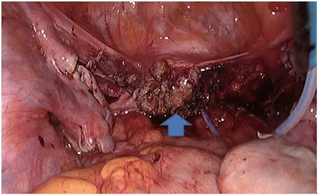
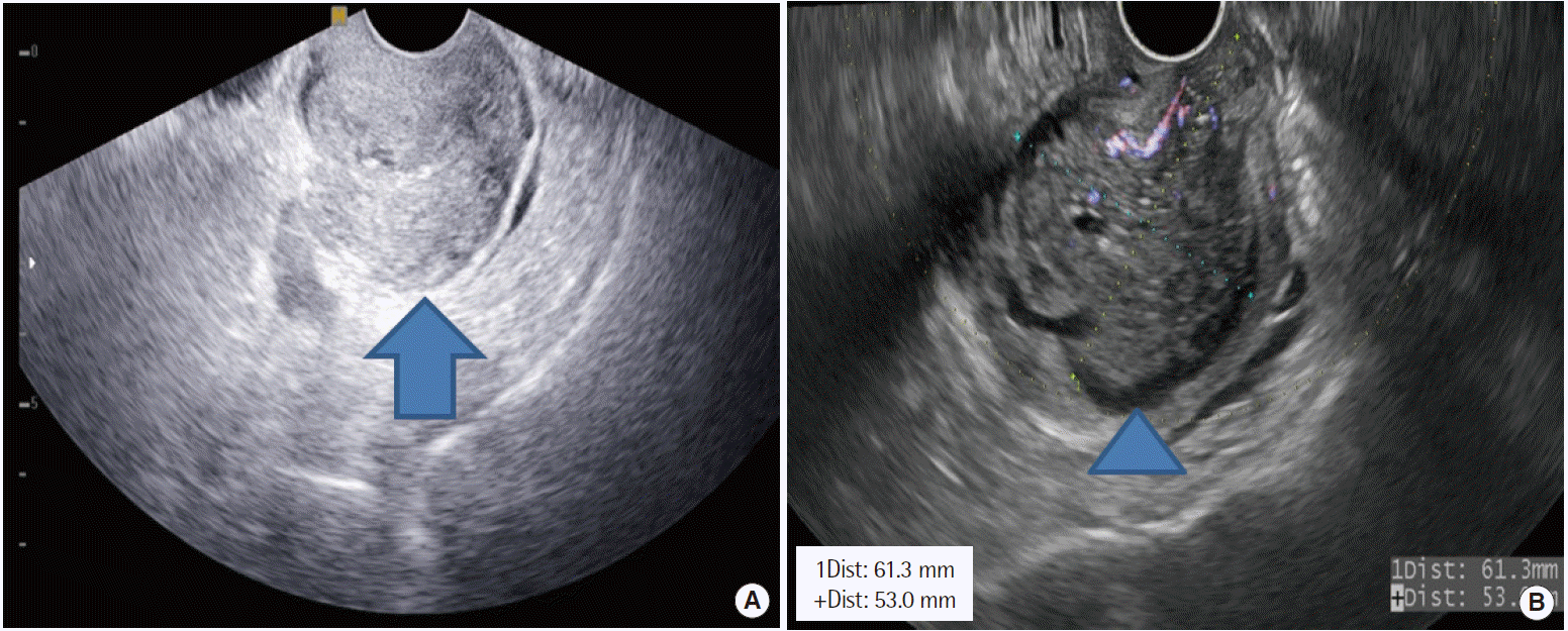
A 40-year-old woman visited emergency clinic of Soonchunnhyang University Cheonan Hospital with a chief complaint of 2-weekhistory of a large amount of yellow and foul odorous vaginal discharge and nausea. The patient also had a fishy vaginal odor in the cervix, which was noticeable following sexual intercourse. The patient had a parity of 2-0-0-2 and a divorced state. In addition, the patient had no notable findings on family and past medical history. Furthermore, the patient also had a 2-month-history of taking supracervical hysterectomy for huge myoma. Preoperatively, the patient had normal Pap smear and very good conditions, except for which the patient had a 5-year-history of undergoing insertion of copper 386 loop. The patient underwent uneventful course without bleeding. Postoperatively, the abdominal cavity was very clear on pelviscopy (Fig. 1). On pelvic ultrasonography, the patient had normal cuff and bilateral ovaries one month postoperatively (Fig. 2A).
On physical examination and serum biochemistry, the patient had a temperature of 36.7˚C, and serum C-reactive protein levels of 1.0 mg/L (normal range, 0.01-3.0 mg/L). The patient had a 61×53 mm abscess on the cuff on transvaginal ultrasonography (Fig. 2B), for which we administered oral antibiotics such as doxycycline and metronidazole. On bacterial culture test with vaginal discharge at that day, gram negative P. bivia was identified. Therefore, the patient was treated with a 14-day course of aerobic specific antibiotics such as cefoxitin plus metronidazole plus doxycycline. One day 14, the size of the abscess on the cuff was decreased to 45×30 mm. Two months thereafter, the patient achieved a complete resolution of the abscess on the cuff without recurrent episodes. In addition, at 12-month follow-up, the patient had no notable findings or recurrences.
DISCUSSION
The DNA of P. bivia has been detected from human vaginal epithelia in 40% of healthy women. The prevalence is relatively higher in women with bacterial vaginosis (BV) [7]. Its detection from the glans penis indicates that it is a causative factor of BV in female sex partners [8]. It is also probable, however, that traumatic events such as human bite might cause P. bivia infections leading to suppurative inguinal adenitis during orogenital sex. It is also a normal oral and vaginal flora as well as a predominant type of bacillus isolated from the respiratory tract infections and their complications, including aspiration pneumonia, lung abscess, chronic otitis media, chronic sinusitis, abscesses in the oral cavity, human bites, brain abscesses, and osteomyelitis. Moreover, P. bivia is the important pathogen in obstetric and gynecologic infections.
P. bivia is present in the digestive tract extending from the oral cavity to the anus [9]. It is considered a common causative agent for infections secondary to animal and human bites. Human and animal bites usually contain polymicrobial, anaerobes that are isolated from more than 2/3 of human and animal bite wound infections, particularly including those associated with abscess formation [10]. There are reports about isolation from the blood of patients delivered by cesarean section as well as those with acute pelvic inflammatory diseases. It has been reported to invade the human cervix and to cause intrauterine infections. It is therefore considered a risk factor of aggravating pregnancy outcomes and fetus development. There is a report suggesting that the concentrations of P. bivia and lipopolysaccharide in the vaginal fluid may contribute to the severity of BV and to the development of adverse pregnancy outcomes, such as preterm labor, premature rupture of membranes, postpartum endometritis, and postoperative pelvic infection [11,12]. In our case, P. bivia in the vagina invade the cervix and caused the postoperative abscess on the cuff. In women with P. bivia undergoing hysterectomy, perioperative treatment with metronidazole eliminates the risk of postoperative cuff infections after hysterectomy [13,14]. In Korea, however, clinicians are not allowed to use the first and second generation cephalosporins for the antimicrobial prophylaxis from immediately before skin incision to postoperative day 1. We could not use additional antibiotics to the patient has no sign of infections.
Our case indicates that clinicians should be aware of possibility of P. bivia infections although rare. And neurologic complications like as brachial plexus injury are rarely associated with laparoscopic procedures [15].
In addition, clinicians should collect anerobic cultures from patients with history of contact to infection sites currently with mucosal damages. Good clinical outcomes are predicted from patients with is positive anerobic gram-negative rods, if they are treated with aerobic specific antibiotics in a timely manner.


