INTRODUCTION
Hypothyroidism is a common disease. The prevalence of hypothyroidism in 2009 for Korea included 248,387 people. Hypothyroidism can be classified as primary hypothyroidism, transient cause and central one. Secondary hypothyroidism refers to a hypothyroid state associated with thyroid stimulating hormone (TSH) deficiency. With the availability of TSH releasing hormone (TRH) assay, it is now possible to separate pituitary origin from hypothalamic hypothyroidism.
Hypothyroidism is mostly caused by primary hypothyroidism. Hashimoto’s thyroiditis is the most common cause of primary hypothyroidism. Secondary hypothyroidism is not common and is usually diagnosed in the context of other anterior pituitary hormone deficiencies, if its origin is from the pituitary gland. Pituitary diseases with multiple anterior pituitary hormone deficiencies usually occur in a predictable sequence, with growth hormone (GH) and gonadotropin deficiency preceding that of TSH and adrenocorticotropin.
However, isolated TSH deficiency is very rare worldwide. It was first reported in 1953 by Shuman [1]. Since then only a few sporadic cases have been reported, and there have been only two reported cases in Korea [2]. We present the case of a Korean female diagnosed with isolated TSH deficiency with a review of the medical literature.
CASE REPORT
A 50-year-old woman with a history of hypothyroidism for 6 months presented to Soonchunhyang University Cheonan Hospital for pain in the right wrist. Radiologic imaging confirmed a distal radius fracture by slip down. She was diagnosed with hypothyroidism during a routine health screen at another facility 6 months prior to presentation. However, despite being recommended to take thyroid hormone, she did not take any of the medication. She had no past history of cardiac or renal disease, and there was no history of significant alcohol intake, smoking or previous surgery. Her family history was also unremarkable. Upon history taking and physical exam, she did not exhibit any manifestation for hypothyroidism.
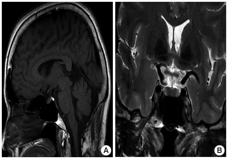
On admission her blood pressure was 120/80 mm Hg with a pulse rate of 80 beats/min. Physical examination was unremarkable and displayed no specific abnormalities besides her radius fracture. Laboratory data showed no abnormal findings except in her thyroid functions: serum white blood cell count was 4,350/μL; hemoglobin 12.8 g/dL; blood urea nitrogen 17.9 mg/dL; creatinine 0.7 mg/dL; serum sodium level 145 mEq/L; serum potassium level 4.1 mEq/L; aspartate aminotransferase 33 IU/L; alanine aminotransferase 23 IU/L; serum glucose 103 mg/dL. Her thyroid function test showed hypothyroidism based on low TSH (0.066 μIU/mL; normal range, 0.27 to 4.2); low free T4 (0.799 ng/dL; normal range, 0.93 to 1.7), and normal T3 (1.03 ng/mL; normal range, 0.8 to 2.0). Antibody to thyroglobulin was 520 IU/mL (normal range, 0 to 115 IU/mL) and the anti-thyroid peroxidase antibody was 600 IU/mL (normal range, 0 to 34 IU/mL). Thyroid ultrasonography showed no abnormailities. These results indicated her hypothyroidism to be of secondary origin. The results of combined anterior pituitary secretion test are shown in Table 1. After administration of exogenous TRH, no TSH response was elicited. However, her prolactin and other pituitary hormone responses after stimulation were normal. Chest X-ray was within normal limits and her electrocardiogram showed a normal sinus rhythm. Pituitary magnetic resonance imaging (MRI) did not show any abnormalities (Fig. 1). The patient was treated with levothyroxine 50 μg per day. After 2 months of treatment, thyroid function returned to normal ranges: TSH 0.046 μIU/mL; free T4 0.946 ng/dL; T3 1.13 ng/mL.
DISCUSSION
Most common causes of primary hypothyroidism are primary, arising from autoimmune thyroiditis and various other iatrogenic causes. Central hypothyroidism, which is from either a pituitary or a hypothalamic origin, is classified as secondary hypothyroidism, which is not a major cause of hypothyroidism.
Pituitary hypothyroidism is characterized by low basal TSH levels in the setting of low free thyroid hormone. Patients with hypothyroidism of hypothalamic origin, on the other hand, may exhibit normal or even slightly elevated TSH levels. The TSH produced in the latter circumstance appears to have reduced biologic activity due to altered glycosylation [3]. Hyposecretion of TSH in pituitary hypothyroidism is ascribed to a reduced mass of functioning thyrotrophs as a consequence of various lesions that can include mechanical compression by tumor, destruction by vascular, inflammatory or physical injuries, alpasia, or hypoplasia. In addition, it may also be idiopathic, when there is no evidence of injury to the pituitary or presence of any neoplastic lesion in the brain. Idiopathic TSH deficiency takes places with correlation to GH deficiency [4], but secretion of other pituitary hormones may also be impaired [5]. Although more common in adults, isolated TSH deficiency also occurs in children and may result in a secondary impairment of GH secretion, including primary deficiency of TSH and GH [6].
Isolated TSH deficiency can occur but is very rare. Several explanations for idiopathic isolated TSH deficiency are possible. Provided that the thyrotrophs are present and morphologically intact, the abnormality could reside in the TRH receptor or at some subsequent step in the transmission of the hypothalamic message, in the process of TSH synthesis, or TSH release. Any abnormalities in the TRH receptor do not involve prolactin-secreting cells because the prolactin response to TRH is normal in these patients [7].
Inherited isolated TSH deficiency is an autosomal recessive disease that results in congenital hypothyroidism and has been reported a few times. It is related to either a single-base substitution or to a nonsense mutation in the TSH-subunit gene, and defects in pituitary specific transcription factor have been also described [8].
The diagnostic criteria for isolated TSH deficiency include 1) a low level of thyroid hormones with low TSH level in the serum, with normal or high serum TRH levels, 2) the absent response of serum TSH to exogenous TRH administration, but with a normal serum prolactin level, and 3) normal secretions of other anterior pituitary hormones. The laboratory data acquired from our patients is compatible with isolated TSH deficiency. The cause of her isolated TSH deficiency was not found in her pituitary MRI. A genetic analysis was not conducted due to her refusal.
The main clinical symptoms of hypothyroidism are fatigue, weak ness, dry skin, feeling cold, hair loss, constipation, dyspnea, hoarseness, and paresthesia. The typical clinical features include a puffy face with edematous eyelids. However, adult patients with isolated TSH deficiency are generally presented with absent or mild symptoms of hypothyroidism [9]. In out patient’s case, she was diagnosed incidentally with hypothyroidism although no symptoms of the condition were shown.
Treatment of TSH deficiency is a daily replacement with levothyroxine. It should be initiated after adequate adrenal function has been established. Dose adjustment is based on thyroid hormone levels and clinical parameters rather than the TSH level [10].
Since the typical symptoms with hypothyroidism are usually lacking, it is clinically important to make the diagnosis of isolated TSH deficiency. Administering thyroid hormone, once the diagnosis has been made, is the only therapy fit for primary hypothyroidism. However, if the patient has an isolated TSH deficiency, it must not be overlooked that the lesion responsible for the disease may lead to other hormonal deficiencies later on. Thus, when a patient has been diagnosed with isolated TSH deficiency, the physician should be alert for any subsequent changes that may require additional evaluation of other hormone levels and the respective therapy for each hormone abnormalities as indicated [6]. We report this case to make note of the fact that both the diagnosis and follow-up care of isolated TSH deficiency must be meticulously done.


