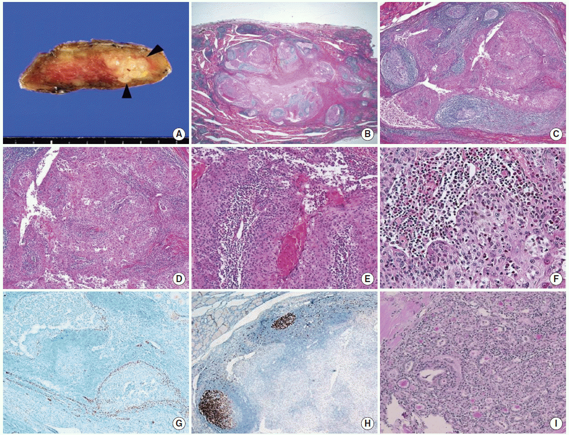
lass='label'>Fig. 3.</div> <div class='caption'> <p>Macroscopic and microscopic findings of the lymphoepithelial cyst of the thyroid gland. (A) Grossly, the specimen has a multinodular, yellowish-gray, solid lesion (arrowhead). (B-F) The lesion is a labyrinth-like cystic lesion lined by squamous epithelial cells with florid lymphoid hyperplasia and focal desquamating keratin. The epithelial components show solid and papillary architecture. Some damaged cellular lesions shows lymphocytic and eosinophilic infiltration (H&E stain; B, × 12.5; C,× 100; D&E,× 200; F, × 400). (G) The p63 is positive in the squamous components (× 200). (H) Ki-67 proliferation index is less than 2% (× 200). (I) Opposite lobe contains a 0.2-cm papillary microcarcinoma, follicular variant (H&E, × 200).</p> <

Download Figure

Fig. 3.

Macroscopic and microscopic findings of the lymphoepithelial cyst of the thyroid gland. (A) Grossly, the specimen has a multinodular, yellowish-gray, solid lesion (arrowhead). (B-F) The lesion is a labyrinth-like cystic lesion lined by squamous epithelial cells with florid lymphoid hyperplasia and focal desquamating keratin. The epithelial components show solid and papillary architecture. Some damaged cellular lesions shows lymphocytic and eosinophilic infiltration (H&E stain; B, × 12.5; C,× 100; D&E,× 200; F, × 400). (G) The p63 is positive in the squamous components (× 200). (H) Ki-67 proliferation index is less than 2% (× 200). (I) Opposite lobe contains a 0.2-cm papillary microcarcinoma, follicular variant (H&E, × 200).